Abstract
Simultaneous acute ischemic stroke (AIS) and acute pulmonary embolism (PE) is an uncommon but life-threatening clinical scenario that presents a significant therapeutic challenge. While AIS requires urgent cerebral reperfusion therapy, PE is primarily managed with anticoagulation or systemic thrombolysis. The coexistence of these conditions creates a difficult balance between preventing neurological deterioration and minimizing the risk of hemorrhagic transformation of cerebral infarction. We report the case of a 68-year-old woman presenting with acute neurological deficits in the setting of suspected pulmonary embolism and discuss the pathophysiology, diagnostic approach, current evidence, and decision-making process regarding thrombolytic therapy. We emphasize that management should be individualized according to stroke severity, PE severity, hemodynamic status, imaging findings, and bleeding risk.
Keywords: Acute ischemic stroke, Pulmonary embolism, Paradoxical embolism, Thrombolysis, Mechanical thrombectomy, Patent foramen ovale.
Introduction
Acute ischemic stroke and pulmonary embolism are individually common medical emergencies. However, their simultaneous occurrence is rare, with only isolated case reports and small case series described in the literature. Their coexistence presents a unique therapeutic dilemma because the recommended treatments may conflict.
Acute ischemic stroke requires rapid reperfusion through intravenous thrombolysis or mechanical thrombectomy, whereas pulmonary embolism is primarily treated with immediate anticoagulation or systemic thrombolysis in high-risk cases. Early anticoagulation after cerebral infarction increases the risk of hemorrhagic transformation, while delaying anticoagulation may worsen pulmonary embolism. Therefore, treatment must be individualized based on the relative severity of both conditions.
Case Presentation
A 68-year-old hypertensive woman presented to the emergency department with sudden-onset confusion and right-sided upper and lower limb weakness of one hour duration.
She reported intermittent episodes of breathlessness over the preceding month. One day before admission, evaluation at another hospital revealed oxygen desaturation to 92% on room air, but no definitive diagnosis was established.
On arrival, she was hemodynamically stable.
Vital signs
| Parameter | Bp | temp | Hear rate | Spo2 |
| value | 140/90 | 98.6 | 112 | 74% in RA |
Neurological examination revealed confusion with right-sided hemiparesis. Her Glasgow Coma Scale score was E4VAM5. Pupils were equal and reactive.
Respiratory examination demonstrated bilateral equal air entry without adventitious sounds.
Cardiovascular examination showed normal heart sounds without murmurs and no elevation of jugular venous pressure.
Investigations
Arterial Blood Gas
| Parameter | Value |
| pH | 7.43 |
| PaCO₂ | 25 mmHg |
| PaO₂ | 42 mmHg* |
| HCO₃ | 35.6 mmol/L |
| Lactate | 0.4 mmol/L |
Echocardiography
- Dilated right atrium
- Dilated right ventricle
- Severe pulmonary hypertension
- Left ventricular ejection fraction approximately 40%
- No pericardial effusion
The echocardiographic findings suggested significant right ventricular pressure overload, raising strong suspicion for pulmonary embolism.
Differential Diagnosis
- Acute ischemic stroke with pulmonary embolism
- Paradoxical embolic stroke through a patent foramen ovale
- Cardioembolic stroke
- Large-vessel atherosclerotic stroke
- Stroke mimic
Discussion
Pathophysiological Link Between Stroke and Pulmonary Embolism
The most plausible mechanism is paradoxical embolism.
A venous thrombus originating in the lower limb travels to the pulmonary circulation, causing pulmonary embolism. Acute obstruction of the pulmonary vasculature increases right ventricular afterload and right atrial pressure. In patients with a patent foramen ovale (PFO), elevated right-sided pressures can reverse the interatrial pressure gradient, allowing thrombus to cross into the systemic circulation and embolize cerebral arteries, producing an acute ischemic stroke.
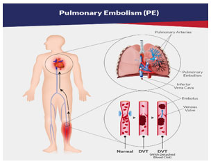
Deep vein thrombosis
↓
Pulmonary embolism
↓
Raised right atrial pressure
↓
Patent foramen ovale opens
↓
Paradoxical embolism
↓
Middle cerebral artery occlusion
↓
Acute ischemic stroke
Other possible mechanisms include atrial fibrillation, left ventricular thrombus following myocardial infarction, infective endocarditis, or severe cardiomyopathy.
Diagnostic Evaluation
Patients with simultaneous neurological deficits and unexplained hypoxemia should undergo parallel evaluation for both stroke and pulmonary embolism.
Stroke Imaging
- Non-contrast CT brain
- CT angiography of head and neck
- CT perfusion (if available)
Pulmonary Embolism Evaluation
- CT pulmonary angiography
- ECG
- Cardiac biomarkers
- Lower-limb venous Doppler
Embolic Source
- Bubble contrast echocardiography
- Continuous ECG monitoring
- Holter monitoring
- Hypercoagulable work-up in selected patients
Therapeutic Dilemma
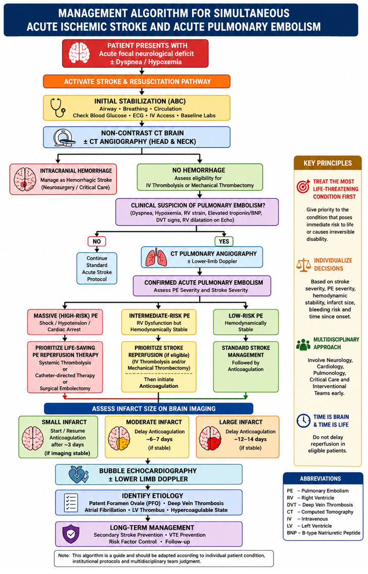
The cornerstone of management is determining which condition poses the greater immediate threat to life or neurological outcome.
| Stroke | Pulmonary embolism |
| Requires immediate reperfusion | Requires anticoagulation |
| Delay reduces neurological recovery | Delay increases mortality |
| Anticoagulation increases hemorrhagic transformation | Thrombolysis may increase intracranial bleeding |
What Determines Whether to Thrombolyse?
The decision to administer thrombolysis is not based solely on the diagnosis of stroke or PE. It depends on four major factors:
-
Stroke Severity
- NIHSS score
- Presence of large vessel occlusion
- Salvageable penumbra
- Time from symptom onset
- Size of infarction
Patients with disabling stroke presenting within the thrombolysis window should generally receive reperfusion therapy if eligible.
-
Pulmonary Embolism Severity
Massive (High-risk) PE
- Shock
- Persistent hypotension
- Cardiac arrest
These patients require immediate reperfusion because mortality is high without treatment.
Intermediate-risk PE
- Right ventricular dysfunction
- Elevated cardiac biomarkers
- Hemodynamic stability
Most patients are treated initially with anticoagulation.
Low-risk PE
Anticoagulation alone is appropriate.
-
Hemodynamic Stability
Hemodynamic status is often the deciding factor.
Stable patients allow time for brain imaging and stroke-directed therapy, whereas unstable patients with massive PE may require immediate reperfusion despite recent cerebral infarction.
-
Risk of Intracranial Hemorrhage
Factors increasing bleeding risk include:
- Large infarct core
- Extensive cerebral edema
- Severe uncontrolled hypertension
- Recent surgery
- Coagulopathy
Practical Management
Scenario 1
Large Vessel Occlusion + Stable PE
- Intravenous thrombolysis if eligible
- Mechanical thrombectomy
- Delay anticoagulation according to infarct size
This approach prioritizes neurological recovery.
Scenario 2
Massive PE + Minor Stroke
Systemic thrombolysis should be prioritized because untreated massive PE carries an immediate risk of death.
Scenario 3
Massive Stroke + Stable PE
Mechanical thrombectomy (if indicated) or intravenous thrombolysis should be performed first. Anticoagulation can be delayed while monitoring for hemorrhagic transformation. Temporary inferior vena cava filter placement may be considered if there is proven proximal DVT and anticoagulation is contraindicated.
Scenario 4
Both Stroke and PE are Life-threatening
Management should involve a multidisciplinary Pulmonary Embolism Response Team (PERT), stroke neurologist, cardiologist, and intensivist. Depending on available expertise, treatment options include systemic thrombolysis, catheter-directed thrombolysis, mechanical thrombectomy, or surgical embolectomy.
Why Is Thrombolysis Based on Severity Rather Than Diagnosis?
The primary objective is to treat the condition with the highest immediate risk of death or irreversible disability.
- A patient with a disabling stroke but stable PE benefits most from urgent cerebral reperfusion.
- A patient with massive PE and minor stroke is more likely to die from circulatory collapse than neurological injury and therefore requires urgent PE-directed reperfusion.
- Stable patients with moderate disease in both vascular territories require individualized management based on imaging, infarct size, and bleeding risk.
Thus, thrombolysis is guided by clinical severity, hemodynamic status, imaging findings, and contraindications—not by the coexistence of stroke and pulmonary embolism alone.
Conclusion
Simultaneous acute ischemic stroke and pulmonary embolism is an uncommon but critical clinical emergency requiring rapid recognition and coordinated multidisciplinary care. The possibility of paradoxical embolism should be considered in patients presenting with acute neurological deficits in association with unexplained hypoxemia or right ventricular strain. Treatment should be individualized after careful assessment of stroke severity, pulmonary embolism severity, hemodynamic stability, infarct size, and bleeding risk. Rather than following a fixed protocol, clinicians should prioritize the condition posing the greatest immediate threat to life or neurological function. Early imaging, timely reperfusion when indicated, and appropriate sequencing of anticoagulation are essential to optimize outcomes.
Suggested References
- 2021 American Heart Association/American Stroke Association Guideline for the Early Management of Patients With Acute Ischemic Stroke.
- 2019 European Society of Cardiology Guidelines for the Diagnosis and Management of Acute Pulmonary Embolism.
- 2024 American College of Chest Physicians Antithrombotic Therapy Guidelines.
- Tintinalli’s Emergency Medicine.
- Rosen’s Emergency Medicine.
This structure is appropriate for submission to an IMA journal as a case report with literature review, and it clearly emphasizes that the decision to thrombolyse depends on clinical severity, imaging, hemodynamic stability, and bleeding risk, rather than the presence of both diagnoses alone.
Mentor

Dr. Ashok nandagopal,
HOD , Department of Emergency Medicine
Kauvery Hospital, Chennai.[1]

Dr. Kowrilakshmi. U,
Department of Emergency Medicine
Kauvery Hospital, Chennai.[1]