Abstract
Introduction: Severely comminuted talar fracture-dislocations are rare in the paediatric population and represent a significant orthopaedic challenge because of the talus’s precarious blood supply and essential role in ankle and hindfoot biomechanics. Associated open injuries and extensive soft tissue damage further complicate management. We report a rare case of a high-energy open Hawkins Type III talar fracture-dislocation with associated calcaneal and medial malleolar fractures, managed using a staged surgical approach.
Case Presentation:
A 14-year-old boy sustained an injury to his left ankle following a fall from the second floor. Clinical examination revealed gross swelling, deformity and a small open wound, classified as Gustilo-Anderson Type I. Computed tomography demonstrated a severely comminuted Hawkins Type III talar fracture-dislocation with disruption of both tibiotalar and subtalar joints, accompanied by comminuted intra-articular calcaneal and displaced medial malleolar fractures. Initial management involved wound exploration, irrigation, debridement and provisional calcaneo-talo-tibial transarticular stabilisation using a Steinmann pin. Following improvement in the soft tissue condition, definitive open reduction and internal fixation was performed. The medial malleolus was fixed using two cannulated cancellous screws, the calcaneal fragments using two Herbert screws and a partially threaded cortical screw, and the talar body using a lateral buttress plate supplemented with two Herbert screws. At four months following definitive fixation, the patient had progressed to weight-bearing.
Conclusion:
Staged surgical management provides a valuable strategy for complex open talar fracture-dislocations, allowing soft tissue recovery before definitive reconstruction while minimising additional vascular compromise. Long-term follow-up remains essential to monitor for avascular necrosis and post-traumatic arthritis.
Keywords: Talar fracture-dislocation; Hawkins Type III; Paediatric trauma; Staged fixation; Open fracture; Calcaneal fracture; Avascular necrosis.
Introduction
Fractures of the talus are uncommon in the paediatric population, and severely comminuted fracture-dislocations involving multiple hindfoot articulations are particularly rare. The talus plays a crucial role in ankle and hindfoot biomechanics, with approximately 60% of its surface covered by articular cartilage and no direct muscular or tendinous attachments. Its predominantly retrograde blood supply, derived from branches of the posterior tibial, anterior tibial and peroneal arteries, makes it particularly vulnerable to vascular compromise following high-energy trauma. Displaced fractures involving the talar body and associated joint dislocations carry a substantial risk of avascular necrosis (AVN), non-union and post-traumatic osteoarthritis. The Hawkins classification describes the severity of talar fracture-dislocations, with Type III injuries characterised by simultaneous dislocation of the tibiotalar and subtalar joints and an associated high risk of AVN. High-energy mechanisms such as falls from height and road traffic accidents may produce extensive talar comminution, frequently accompanied by calcaneal and malleolar fractures. The presence of an open wound and extensive soft tissue injury further increases the risk of infection and wound-related complications. Management requires a careful balance between anatomical reconstruction and preservation of the remaining vascular supply and soft tissue envelope. In injuries associated with significant swelling and open wounds, immediate definitive internal fixation may increase the risk of wound complications. A staged approach involving initial wound debridement and provisional stabilisation, followed by definitive fixation after soft tissue recovery, is therefore an important treatment strategy. We present a rare case of a severely comminuted open Hawkins Type III talar fracture-dislocation with associated intra-articular calcaneal and medial malleolar fractures in a 14-year-old boy, highlighting the role of staged reconstruction in managing complex paediatric hindfoot injuries.
Case Report
A 14-year-old boy presented to the Emergency Department following an alleged fall from the second floor, sustaining an injury to his left ankle. Clinical examination revealed gross swelling and deformity of the left ankle and hindfoot, with a small breach in skin integrity consistent with a Gustilo-Anderson Type I open injury. Distal neurovascular status was assessed, and the affected limb was immobilised with a splint. Computed tomography of the left ankle and hindfoot demonstrated a severely comminuted fracture-dislocation of the talus with complete disruption of the talar body and gross displacement of multiple fragments, resulting in marked incongruity of the tibiotalar and subtalar joints. Based on the simultaneous dislocation of both articulations, the injury was classified as a Hawkins Type III fracture-dislocation. Associated injuries included a comminuted intra-articular calcaneal fracture involving the lateral process, sinus tarsi, peroneal tubercle and anterior process, with extension into the subtalar and calcaneocuboid articular surfaces. A comminuted displaced medial malleolar fracture was also identified. Extensive periarticular soft tissue oedema and multiple intra-articular and intramuscular air locules were noted, consistent with the open nature of the injury. Considering the extensive osseous comminution, open wound and significant soft tissue compromise, a staged surgical approach was planned.
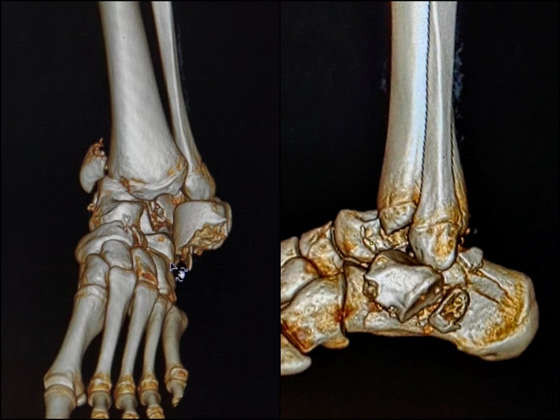
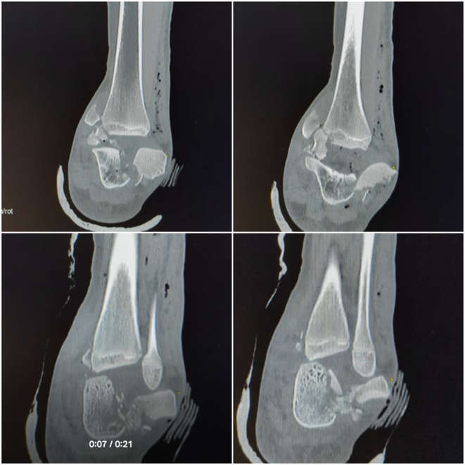
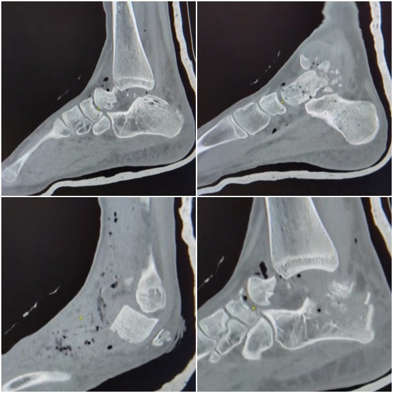
Stage I: Emergency Wound Debridement and Provisional Stabilisation
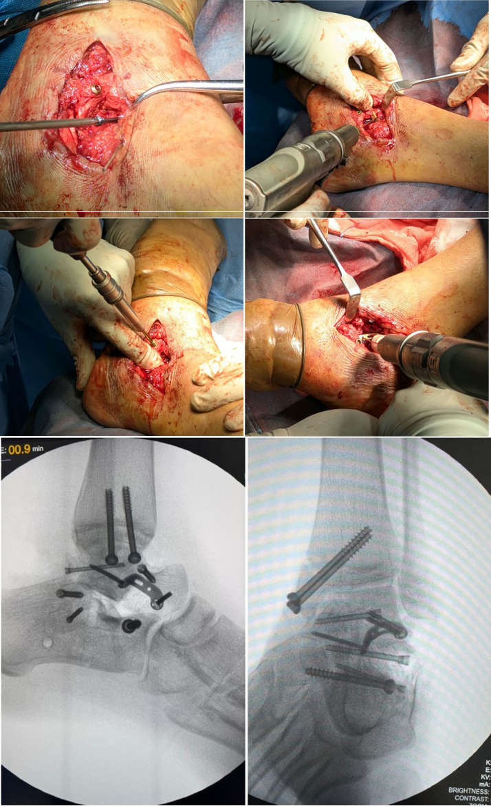
The initial procedure involved thorough wound exploration, irrigation and surgical debridement in accordance with open fracture management principles. Following debridement, provisional stabilisation of the ankle and hindfoot was achieved using a calcaneo-talo-tibial transarticular Steinmann pin under fluoroscopic guidance. The pin spanned the tibiotalar and subtalar joints, maintaining limb length, alignment and rotation while providing temporary stability to the severely disrupted hindfoot. This provisional stabilisation prevented further displacement of fracture fragments, reduced movement at the injured articulations and protected the surrounding soft tissue envelope. The staged approach allowed adequate time for soft tissue recovery before definitive fixation, thereby reducing the potential risk of wound breakdown, infection and complications associated with operating through compromised tissues.
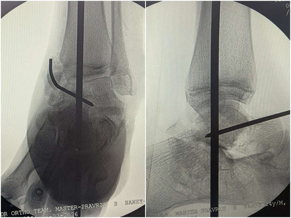
Stage II: Definitive Anatomical Reconstruction and Internal Fixation
Following improvement in the local soft tissue condition, the patient underwent the second stage of surgery for definitive open reduction and internal fixation. The primary objectives were restoration of anatomical alignment, reconstruction of the articular surfaces, stable fixation and preservation of the residual vascular supply to the talar fragments. The medial malleolar fracture was reduced and fixed using two cannulated cancellous screws. The calcaneal fragments were stabilised using two Herbert screws and one partially threaded cortical screw. Anatomical reduction of the severely comminuted talar body was achieved and secured using a lateral buttress plate supplemented with two Herbert screws. Particular care was taken to minimise periosteal stripping and unnecessary soft tissue dissection around the talus to preserve its remaining extraosseous vascular contributions. Herbert screws provided interfragmentary compression while minimising hardware prominence near the articular surfaces. Immediate postoperative fluoroscopic imaging demonstrated satisfactory restoration of talar height and contour, improved hindfoot alignment and congruity of the tibiotalar and subtalar joints. The positions of the medial malleolar screws, calcaneal screw and talar plate and screw construct were satisfactory.
Postoperative Management and Follow-up
Postoperatively, the limb was immobilised, and the patient was maintained on a strict non-weight-bearing protocol to protect the fixation and minimise stress on the tenuous vascular supply of the talus. Regular wound surveillance and appropriate antibiotic management were undertaken in accordance with open fracture protocols. Rehabilitation was planned according to clinical progress and radiological evidence of fracture healing. At four months following the second-stage definitive fixation, the patient had progressed to weight-bearing, representing an encouraging early functional milestone following reconstruction of the severely comminuted talar, calcaneal and medial malleolar fractures. Continued clinical and radiological follow-up is essential to assess fracture union, talar viability, ankle and subtalar range of motion and the development of late complications, particularly avascular necrosis and post-traumatic osteoarthritis.
Discussion
Severely comminuted talar fracture-dislocations are among the most challenging injuries in orthopaedic trauma because of the complex anatomy, limited vascular supply and essential biomechanical role of the talus. Their occurrence in the paediatric and adolescent population is particularly rare. The present case represents a high-energy injury involving three bones of the hindfoot, namely the talus, calcaneus and medial malleolus, accompanied by an open wound and extensive soft tissue compromise. The principal challenge was achieving anatomical reconstruction while protecting the compromised soft tissue envelope and preserving the remaining blood supply to the talus. Hawkins Type III injuries involve complete dislocation of both the tibiotalar and subtalar joints and are associated with a particularly high risk of AVN. Adult literature frequently reports AVN rates of approximately 70–90% in Type III injuries, although these estimates should be interpreted cautiously when applied to adolescent patients. The most important aspect of management in this case was the decision to adopt a staged surgical approach. In high-energy open fractures with extensive periarticular swelling, immediate definitive internal fixation may necessitate extensive dissection through compromised soft tissues, increasing the risk of wound breakdown, infection and further damage to the vascular supply. Therefore, appropriate soft tissue management is as important as anatomical fracture reduction in determining the eventual outcome. The first stage focused on wound management and temporary skeletal stabilisation. Thorough irrigation and debridement were essential to remove contaminated and devitalised tissue and reduce the risk of infection. Provisional calcaneo-talo-tibial transarticular pin fixation maintained the alignment of the ankle and hindfoot while limiting further movement at the fracture sites. Importantly, temporary stabilisation allowed the soft tissue envelope to recover before definitive reconstruction. This approach follows the principles of damage-control orthopaedics, prioritising early injury control and soft tissue protection before definitive fixation.The timing of the second stage is determined by wound healing and local soft tissue recovery. Definitive fixation should be undertaken when the tissues are sufficiently recovered to tolerate further surgical intervention and satisfactory wound closure can be achieved. Proceeding too early through markedly swollen or compromised tissues may increase the likelihood of wound dehiscence, infection and implant-related complications. The second stage involved anatomical reconstruction of the talus, calcaneus and medial malleolus. Restoration of talar height, contour and articular congruity was essential to re-establish the relationship between the tibia, talus and calcaneus and preserve the possibility of functional ankle and subtalar movement. The combination of a lateral buttress plate and Herbert screws provided fixation of the comminuted talar body, while the associated calcaneal and medial malleolar fractures were stabilised using appropriate screw constructs. Careful handling of the fragments and minimisation of periosteal stripping were important to avoid further vascular injury. The patient’s age of 14 years is also relevant. Although growth-related complications are important considerations in paediatric fractures, the principal long-term concerns in this case are vascular viability of the talus, maintenance of joint congruity, fracture union and development of post-traumatic osteoarthritis. The postoperative course provides an encouraging early functional outcome. Progression to weight-bearing at four months following definitive fixation is a positive milestone. However, weight-bearing alone does not establish complete fracture union or exclude avascular necrosis. AVN, non-union, infection, post-traumatic arthrosis of the tibiotalar and subtalar joints and restricted joint mobility remain important potential complications. The Hawkins sign, characterised by subchondral radiolucency in the talar dome, is an important radiographic indicator of preserved talar vascularity and is generally assessed approximately 6–8 weeks following injury. Serial radiographic evaluation is essential to monitor fracture union and identify early features of osteonecrosis. Long-term follow-up should include assessment of pain, wound condition, ankle and subtalar range of motion, weight-bearing tolerance and functional mobilisation. Patients developing symptomatic AVN or advanced post-traumatic arthritis may eventually require additional procedures, including arthrodesis. Continued surveillance is therefore essential despite the encouraging progress observed at four months.
Conclusion
Severely comminuted open Hawkins Type III talar fracture-dislocations with associated calcaneal and medial malleolar fractures are exceptionally rare and challenging injuries in the adolescent population. Successful management requires anatomical reconstruction, meticulous wound care, protection of the soft tissue envelope and preservation of residual talar vascularity. This case highlights the importance of a staged surgical strategy involving initial wound debridement and provisional calcaneo-talo-tibial transarticular stabilisation, followed by definitive internal fixation after adequate soft tissue recovery. Progression to weight-bearing at four months represents an encouraging early functional outcome. Nevertheless, long-term clinical and radiological follow-up remains essential to monitor fracture union, avascular necrosis, post-traumatic arthritis and functional recovery.
References
- Parekh SG, Kadakia RJ. Avascular necrosis of the talus. J Am Acad Orthop Surg. 2021;29(6):e267-78. doi:10.5435/JAAOS-D-20-00418.
- Alley MC, Vallier HA, Tornetta P 3rd; Orthopaedic Trauma Research Consortium. Identifying risk factors for osteonecrosis after talar fracture. J Orthop Trauma. 2024;38(1):25-30. doi:10.1097/BOT.0000000000002706.
- Griffin JT, Landy DC, Mechas CA, Nazal MR, Foster JA, Moghadamian ES, et al. The Hawkins sign of the talus: the impact of patient factors on prediction accuracy. J Bone Joint Surg Am. 2024;106(11):958-65. doi:10.2106/JBJS.23.00906.
- Waseem S, Nayar SK, Vemulapalli K. Paediatric talus fractures: a guide to management based on a review of the literature. Injury. 2022;53(3):1029-37. doi:10.1016/j.injury.2021.12.024.
- Lee GW, Lee KB. Current management of talar fractures. J Musculoskelet Trauma. 2022;35(1):31-7. doi:10.12671/jkfs.2022.35.1.31.

Dr. Asim Chundeli,
DNB Resident, Orthopedics
Kauvery Hospital, Chennai.[1]

Dr. A. William Abraham,
Consultant Orthopedic Surgeon
Kauvery Hospital, Chennai.[1]