INTRODUCTION
Aortic intramural hematoma (IMH) is an acute aortic syndrome caused by hemorrhage within the aortic media, in the absence of a conventional patent false lumen. IMH may progress to aortic dissection, rupture, aortic enlargement, or branch-vessel malperfusion. Malperfusion and persistent or recurrent pain are recognized features of complicated IMH and are associated with increased morbidity and mortality. [1]
Large-vessel vasculitis (LVV) encompasses inflammatory disorders affecting the aorta and its major branches, principally Takayasu arteritis (TAK) and giant cell arteritis. Takayasu arteritis predominantly affects younger individuals and may involve the aorta, its major branches, and the renal or mesenteric arteries. Vascular imaging, including computed tomography angiography (CTA), magnetic resonance imaging, ultrasonography, and ^18F-fluorodeoxyglucose positron emission tomography (FDG-PET), has an important role in identifying mural inflammation and defining vascular involvement. [2–4]
The coexistence of active large-vessel inflammation and acute aortic IMH presents a diagnostic challenge because fever, leukocytosis, markedly elevated inflammatory markers, abdominal pain, and renal dysfunction may initially suggest an infectious or renal process. This case illustrates extensive acute IMH with severe unilateral renal malperfusion in a 39-year-old man, initially managed as probable pyelonephritis, in whom subsequent vascular imaging and FDG-PET/CT demonstrated extensive aortic disease and active inflammation involving multiple major arterial branches.
Case Presentation
A 39-year-old man was admitted with a 1-day history of high-grade fever, headache, severe abdominal pain radiating to the back, and left flank pain with tenderness. There was no history of chills or rigors. He had a background history of poorly controlled systemic hypertension for approximately 4 years, with multiple previous episodes of elevated blood pressure/accelerated hypertension. His previous antihypertensive treatment had included calcium-channel blockers, alpha-blockers, nitroglycerin, and other antihypertensive therapy.
On admission, his blood pressure was 160/110 mmHg and pulse rate was 126/min. Temperature was documented as 100.3°F. Oxygen saturation was 98% on room air. He was conscious and oriented. Respiratory examination revealed bilateral air entry, and cardiovascular examination revealed S1 and S2. The abdomen was soft with left flank tenderness and abdominal pain radiating to the back. Central nervous system examination was documented as normal.
An outside abdominal ultrasound had demonstrated gallbladder sludge and a gallbladder polyp. An outside CT abdomen was reported as mildly bulky pancreas, otherwise normal. Initial laboratory investigations revealed leukocytosis with a total leukocyte count of 18,700/µL, with serum creatinine of 1.06 mg/dL. Liver function tests and pancreatic enzymes were normal.
In view of fever, leukocytosis, left flank tenderness, and perinephric inflammatory changes subsequently noted on CT, acute pyelonephritis was initially considered and the patient was started on IV antibiotic cefoperazone-sulbactam. However, urine examination demonstrated no significant pyuria, urine and blood cultures showed no growth, and abdominal/flank pain persisted. Leukocytosis and systemic inflammation also persisted despite 3 days of antimicrobial therapy. The total leukocyte count remained approximately 19,430/µL and CRP remained markedly elevated at approximately 266–291 mg/L.
Renal function subsequently deteriorated, with serum creatinine increasing from 1.06 to 1.67 mg/dL and urea reaching 40.3 mg/dL. Given the persistent abdominal pain, severe hypertension, and worsening renal function, renal vascular evaluation was performed. Renal artery Doppler demonstrated abnormal high-resistance flow in the left renal vascular bed, with reduced peak systolic velocity in the left renal artery, increased resistive index, and reversal of diastolic flow in the hilar, segmental, interlobar, and arcuate branches. Vascular surgeon opinion was obtained and advised for a CT aortogram.
A CT aortogram was subsequently performed after obtaining high risk consent in view of elevated renal parameters and the possibility of contrast induced nephropathy (CIN). CT Aortogram demonstrated extensive aortic intramural hematoma involving the aortic arch, descending thoracic aorta, and abdominal aorta, extending from immediately distal to the right innominate artery origin to above the aortic bifurcation. Multiple focal intramural blood pools/ulcer-like projections were identified, several communicating with the aortic lumen. There was associated involvement of the proximal left internal carotid and left subclavian arteries and the ostial/proximal celiac and superior mesenteric arteries, without significant luminal stenosis.
The most significant complication was severe left renal malperfusion, with non-opacification of the left main renal artery, minimal residual upper-pole segmental arterial supply, absent arterial-phase renal enhancement, and absent left renal contrast excretion, concerning for severe left renal ischemia/infarction. No frank aortic rupture or large periaortic hematoma was identified. The radiological impression was an extensive arch-origin intramural hematoma categorized as Stanford type A IMH with extensive distal propagation and severe left renal malperfusion.
Vascular surgeon review obtained and advised for conservative management and to start anti-inflammatory therapy with IV Steroids. Because of the patient’s young age, extensive aortic and branch-vessel involvement, marked inflammatory response, and concern for an underlying inflammatory aortopathy, further evaluation for large-vessel vasculitis was undertaken. Rheumatologist opinion was obtained and advised for FDG PET CT to demonstrate the inflammatory aetiology and to diagnose vasculitis.
FDG-PET/CT demonstrated diffuse increased FDG uptake along the aortic wall involving the ascending aorta, aortic arch, descending thoracic aorta, and abdominal aorta, with additional uptake involving the brachiocephalic trunk and origins of the right subclavian and common carotid arteries. Patchy increased uptake was also noted along the abdominal aortic wall and at the origins of the superior mesenteric artery, celiac artery, and left renal artery. These findings were reported as suggestive of active vasculitis. The PET-CT also demonstrated the crescentic mural abnormality corresponding to the previously demonstrated extensive aortic IMH.
Further autoimmune evaluation showed negative C-ANCA and P-ANCA, a negative ANA profile, normal C3 and C4, non-reactive serology, and negative RPR. Antiphospholipid testing was negative, including beta-2 glycoprotein antibodies, anticardiolipin antibodies, and lupus anticoagulant. Factor V Leiden mutation analysis did not detect the G1691A mutation.
An ophthalmological evaluation was performed because of the history of severe hypertension. Fundus examination demonstrated attenuation of the arterioles and arteriovenous crossing changes, with grade 2 hypertensive retinopathy. The macula was normal and there was no evidence of papilledema.
Vascular surgery advised strict blood pressure control and conservative management, with no surgical intervention at that time. Rheumatology considered the findings consistent with undifferentiated large-vessel vasculitis, with follow-up for possible Takayasu arteritis.
The patient received intravenous methylprednisolone 500 mg daily for 3 days, followed by oral prednisolone at 0.75 mg/kg/day (60 mg once daily). CRP gradually decreased following initiation of corticosteroid therapy. He was discharged in clinically stable condition and advised for outpatient follow-up.
INVESTIGATIONS
Laboratory investigations
| Investigation | Result |
| Hemoglobin, initial | 14.1 g/dL |
| Total leukocyte count, initial | 18,700/µL |
| Creatinine, initial | 1.06 mg/dL |
| HbA1c | 5.3% |
| Thyroid function tests | Normal |
| Liver function tests | Within normal limits |
| Lipase | Normal |
| Serum cortisol | 29 |
| Overnight dexamethasone suppression
test |
Normal |
| CRP | ~266–291 mg/L, Reduced to 90 mg/dl
after IV Steroids. |
| Creatinine at discharge | 1.42 mg/dL |
| Urea | 40.3 mg/dL |
| Homocysteine | 6.1 |
| C and P-ANCA | Negative |
| ANA profile | Negative |
| C3/C4 | Normal |
| Serology | Non-reactive |
| RPR | Negative |
Urinalysis- No significant pyuria, RBC: 4–6/HPF, Blood: 1+, Protein: Trace
Microbiology
Endocrine evaluation
Background of uncontrolled hypertension and a tiny 3 × 3 mm lipid-rich right adrenal cortical adenoma was identified on CT.
The available biochemical evaluation did not support pheochromocytoma or autonomous cortisol secretion.
Imaging
Renal artery Doppler
The examination showed:
The differential included medical renal disease and renal vein thrombosis.
CT aortogram
The CT demonstrated:
Extensive arch-origin intramural hematoma categorized as Stanford type A IMH with extensive distal propagation, complicated by severe left renal malperfusion.
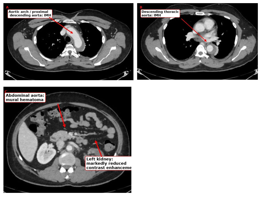
Figure 1 CT aortogram demonstrating extensive aortic intramural hematoma with left renal malperfusion. (A) Intramural hematoma involving the aortic arch and proximal descending thoracic aorta. (B) Extensive mural hematoma involving the descending thoracic aorta.
FDG-PET/CT
FDG-PET/CT showed:
Suggestive of active vasculitis.
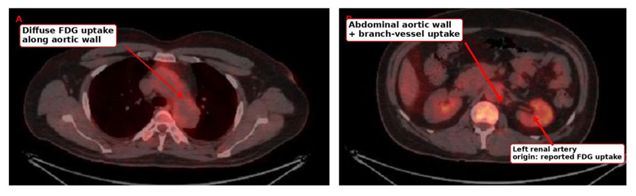
Figure 2 FDG PET-CT demonstrating metabolically active large-vessel inflammation. (A) Increased FDG uptake along the thoracic aortic wall. (B) Increased FDG uptake involving the abdominal aortic wall and proximal branch-vessel regions, including the origins of the celiac artery, superior mesenteric artery and left renal artery.
DIFFERENTIAL DIAGNOSIS
– Acute pyelonephritis
Acute pyelonephritis was the initial working diagnosis because of fever, leukocytosis, flank tenderness, and perinephric inflammatory changes. However, there was no significant pyuria, urine culture showed no growth, and blood culture was negative. Furthermore, abdominal/flank pain, leukocytosis, and markedly elevated CRP persisted despite 3 days of cefoperazone-sulbactam. Subsequent CT angiography demonstrated extensive aortic disease with severe left renal malperfusion, providing an alternative explanation for the clinical syndrome.
– Acute pancreatitis
Abdominal pain radiating to the back and an outside CT report of a mildly bulky pancreas prompted consideration of pancreatitis. However, serum amylase and lipase were normal, and the subsequent vascular imaging demonstrated a significant alternative pathology.
– Pheochromocytoma
Severe hypertension, headache, abdominal pain, and an adrenal lesion prompted biochemical evaluation. However, the adrenal lesion was only approximately 3 × 3 mm and lipid-rich. The available urinary metanephrine was not elevated, and overnight dexamethasone suppression was normal. The findings therefore did not support a functional adrenal tumour as the cause of the current presentation.
– Renal vascular ischemia/infarction
The combination of severe flank pain, microscopic hematuria, worsening renal function, abnormal renal Doppler findings, and subsequent non-opacification of the left renal artery strongly supported severe left renal arterial malperfusion with probable renal ischemia/infarction.
– Renal vein thrombosis
Renal vein thrombosis was included in the initial Doppler differential because of the high-resistance intrarenal waveform and reversal of diastolic flow. However, CT angiography subsequently demonstrated non-opacification of the left main renal artery and absent renal enhancement/excretion in the setting of extensive aortic IMH, providing stronger evidence for arterial malperfusion.
– Acute aortic syndrome
The CT aortogram established an extensive aortic IMH involving the arch, descending thoracic and abdominal aorta. The associated severe left renal malperfusion constituted a major complication. Multiple intramural blood pools/ulcer-like projections and branch-vessel involvement further characterized the acute aortic process.
– Large-vessel vasculitis
Large-vessel vasculitis became a major consideration because of the patient’s age, severe inflammatory response, extensive aortic involvement, branch-vessel abnormalities, and FDG-PET/CT evidence of active inflammation involving the aorta and multiple major arterial branches.
Takayasu arteritis was specifically considered because of the patient’s age and vascular distribution. However, the treating rheumatology assessment described the condition as undifferentiated large-vessel vasculitis, with follow-up for possible Takayasu arteritis, and the available data do not establish a definitive Takayasu diagnosis.
The negative ANCA, ANA, normal complement levels, and negative antiphospholipid testing did not identify an alternative systemic autoimmune disorder. These negative results do not by themselves exclude Takayasu arteritis.
FINAL DIAGNOSIS
-
Active undifferentiated large-vessel vasculitis/aortitis, highly suspicious for Takayasu arteritis
Supported by:
-
Complicated extensive acute aortic intramural hematoma (IMH), radiologically categorized as Stanford type A, with extensive distal propagation
Complicated by: Severe left renal arterial malperfusion with probable left renal ischemia/infarction.
TREATMENT GIVEN
The patient was initially treated for probable acute pyelonephritis with: Cefoperazone-sulbactam for 3 days. In view of persistent abdominal/flank pain, severe hypertension, inflammatory response, and deterioration in renal function, further vascular evaluation was performed and the diagnosis of extensive acute aortic IMH with severe left renal malperfusion was established.
Vascular surgery opinion was obtained. Strict blood pressure control and conservative management were advised, with no surgical intervention at that time. Rheumatology opinion was obtained and the condition was considered undifferentiated large-vessel vasculitis, with follow-up for possible Takayasu arteritis.
The patient was treated with intravenous methylprednisolone 500 mg daily for 3 days followed by Oral prednisolone 60 mg once daily (0.75 mg/kg/day). CRP was documented to gradually decrease after initiation of corticosteroid therapy. At discharge, the patient was clinically stable.
LEARNING POINTS
- Acute aortic syndromes should remain in the differential diagnosis of severe abdominal or flank pain in patients with marked hypertension, particularly when pain radiates to the back and renal function deteriorates.
- Fever, leukocytosis, and markedly elevated CRP can accompany acute aortic and ischemic vascular disease and may mimic infection. In this case, persistent inflammation despite antimicrobial therapy and the absence of significant pyuria or microbiological growth prompted reassessment of the initial diagnosis.
- FDG-PET/CT can demonstrate active large-vessel inflammation extending beyond the anatomical region of an acute aortic lesion. In this patient, FDG uptake involved the aorta and multiple major arterial branches, supporting an active large-vessel inflammatory process. Imaging should nevertheless be interpreted in conjunction with the clinical and anatomic findings because FDG uptake is not specific for a particular vasculitis. [2–4]
- In a young patient with extensive aortic and branch-vessel involvement, an underlying large-vessel vasculitis should be considered, particularly Takayasu arteritis. However, the distinction between confirmed Takayasu arteritis and undifferentiated large-vessel vasculitis requires integration of clinical examination, vascular imaging, and longitudinal follow-up.
- A small adrenal incidentaloma should not distract from a demonstrated vascular cause of severe hypertension. In this case, the 3 × 3 mm lipid-rich adrenal lesion had no supportive biochemical evidence of a functional adrenal tumour.
REFERENCES
- Isselbacher EM, Preventza O, Hamilton Black J 3rd, Augoustides JG, Beck AW, Bolen MA, et al. 2022 ACC/AHA Guideline for the Diagnosis and Management of Aortic Disease: A Report of the American Heart Association/American College of Cardiology Joint Committee on Clinical Practice Guidelines. 2022;146:e334-e482. doi:10.1161/CIR.0000000000001106. (AHA Journals)[1]
- Maz M, Chung SA, Abril A, Langford CA, Gorelik M, Guyatt G, et 2021 American College of Rheumatology/Vasculitis Foundation Guideline for the Management of Giant Cell Arteritis and Takayasu Arteritis. Arthritis Rheumatol. 2021;73(8):1349-1365. doi:10.1002/art.41774. (PubMed)[2]
- Dejaco C, Ramiro S, Bond M, Bosch P, Ponte C, Mackie SL, et al. EULAR recommendations for the use of imaging in large vessel vasculitis in clinical practice: 2023 update. Ann Rheum Dis. 2024;83(6):741-751. doi:10.1136/ard-2023-224543. (Annals of the Rheumatic Diseases)[3]
- Bosch P, Bond M, Dejaco C, Ponte C, Mackie SL, Falzon L, et Imaging in diagnosis, monitoring and outcome prediction of large vessel vasculitis: a systematic literature review and meta-analysis informing the 2023 update of the EULAR recommendations. RMD Open. 2023;9(3):e003379. doi:10.1136/rmdopen-2023-003379. (PubMed)[4]
- Marla R, Migrino R, Osipov V, Lilly RE. Takayasu arteritis presenting as type A intramural hematoma—an unusual presentation. Int J Cardiol. 2010;145(1):e30-e32. doi:10.1016/j.ijcard.2008.12.132. (PubMed)[5]
- Mukhtyar CB, Nikiphorou E, Bartoletti A, Dey M, Ducker G, Misra DP, et al. 2025 EULAR recommendations for the management of polymyalgia rheumatica and primary large vessel vasculitis. Ann Rheum Dis. Published online July 21, 2026. doi:10.1016/j.ard.2026.06.009. (PubMed)[6]

Dr Vignesh A S
DNB Medicine Resident
Kauvery Hospital, Chennai.[7]

Dr Sivaram Kannan
Chief Consultant Internal Medicine
Kauvery Hospital, Chennai.[7]