
How Heart Scans Are Helping Athletes Stay Safe While Staying in the Game
As more people — from weekend joggers, fitness enthusiasts to professional athletes — push their limits in sports and fitness, a new medical speciality is stepping into the spotlight: sports cardiology. This field looks at how exercise affects the heart, and how doctors can tell the difference between healthy changes from training and dangerous heart disease.
Why does this matter? Because sometimes the heart of a highly trained athlete can look abnormal on tests — even when it’s perfectly healthy. On the other hand, a hidden heart problem might be mistaken for a harmless “athlete’s heart”. Both mistakes can have serious consequences – unnecessarily ending an athlete’s career, or worse, missing a life-threatening condition.
The Role of Cardiac Imaging/Radiology
Modern imaging tests — especially cardiac MRI (CMR) and coronary CT angiography (CCTA) — have become powerful tools in solving this puzzle.
- Cardiac MRI can show detailed pictures of the heart’s structure and even the tissue itself. This helps doctors see whether changes in size, wall thickness, or function are simply from training or signs of disease.
- Coronary CT angiography takes high-resolution pictures of the heart’s blood vessels, helping detect rare artery problems in young athletes or early signs of heart disease in older athletes.
These scans don’t just help diagnose problems — they also guide decisions about whether it’s safe for someone to compete, when to return after an illness or heart event, and how to monitor long-term heart health.
Training Changes vs. True Disease
Endurance training (like marathon running) and resistance training (like weightlifting) remodel the heart in different ways. Radiologists and cardiologists must know what’s “normal for an athlete” so they don’t confuse it with a dangerous condition. For example:
- Mild thickening of the heart muscle and slightly lower pumping function may be a normal adaptation.
- In other contexts, the same findings could mean cardiomyopathy, a serious disease.
That’s why imaging is interpreted alongside history, ECGs, exercise tests, and the type of sport someone plays.
When Do Athletes Need These Scans?
Not every athlete requires advanced imaging. Doctors usually recommend scans if:
- There are concerning symptoms, such as chest pain, fainting, or palpitations.
- Routine tests like ECG or echocardiogram raise questions.
- The athlete is recovering from a heart condition or event.
This ensures scans are used where they matter most — helping those truly at risk.
A Team Effort
Sports cardiology is growing fast. Radiologists, cardiologists, team doctors, and trainers all work together to keep athletes safe. It’s not just about reading scans — it’s about putting results into the bigger picture of each person’s health and sport.
Takeaway for Athletes and Families
- Most changes seen in an athlete’s heart are normal adaptations to training.
- Advanced imaging like CMR and CCTA can be lifesaving when used in the right situations.
- If you’re an athlete with unexplained symptoms (chest pain, fainting, palpitations, etc.), talk to your doctor/cardiologist. Early and accurate evaluation — including imaging if needed — can mean the difference between safe play and serious risk.
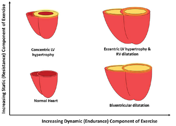
This illustration shows how the heart adapts to different types of exercise. Exercises with more strength training (static) tend to make the heart muscle thicker. Exercises with more endurance training (dynamic) tend to make the heart chambers larger, especially the right side. When both strength and endurance training are intense, the heart shows both thicker muscle and a larger chamber.
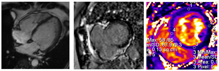
Arrhythmogenic Cardiomyopathy in a Young Athlete
In a cardiac MRI of a 20-year-old volleyball/basketball player with a family history of heart disease, the right side of the heart was enlarged and weak, with poor pumping function. Six months later, follow-up scans showed early scarring of the heart muscle. Although she had no symptoms at first, she later developed ventricular arrhythmia, a heart rhythm problem. She eventually required a defibrillator device to restore a normal heartbeat, and later a heart transplant.
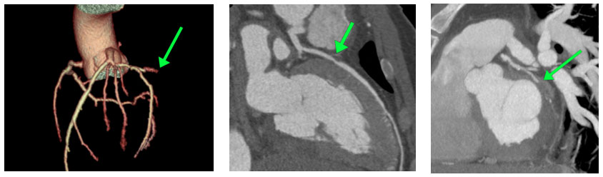
Heart Artery Blockage in a 40-Year-Old Runner
A long-distance runner came to the emergency department with sudden chest pain. Blood tests showed elevated troponin, a marker of heart injury, with no other significant medical/family history. Coronary CT angiography revealed a severe (70%) narrowing in the left anterior descending artery and a complete blockage in the left circumflex artery. The patient immediately underwent stent placement by the cardiologist, restoring normal blood flow and thus averting a life‑threatening situation.
Sports cardiology reminds us: With imaging, we can protect the hearts that sport has trained.

Dr Asik Ali
Consultant Radiologist,
Kauvery Hospital, Chennai
Courtesy: Radiographics 2025
