A 40-year-old male, known case of Ca lung with metastases to ribs and adrenal, was admitted with the complaints of breathing difficulty. Blood investigations were within normal limits, and chest X-ray showed moderate right-sided pleural effusion. Hence, this patient was posted for thoracoscopy and pleural biopsy.
PREOP EVALUATION
HR 98/min, BP – 110/60 mmHg, RR 22/min, Spo2 98% RA
Blood investigations were within normal range
600 ml of pleural fluid was drained previous day for better intraoperative status
Considering the respiratory concerns, thoracoscopy was planned under Serratus Anterior Plane Block to maintain respiratory stability.
SERRATUS ANTERIOR PLANE BLOCK
ANATOMY
Serratus anterior arises from lateral aspect of 1st to 8th rib and inserts in the medial border of scapula
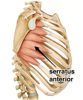
Two spaces
- Between latissimus dorsi and serratus anterior
- Between serratus anterior and external intercostal muscle
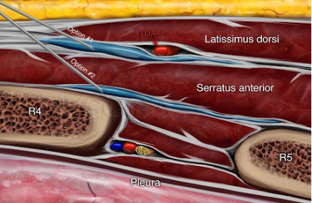
Pictures from Nysora.com
SABP aims to target nerves such as
- Lateral cutaneous branches of intercostal nerves (T3–T9)– originates from anterior rami of thoracic spinal region, supplies sensory innervation to the skin
- Long thoracic nerve– originates from C5, C6, C7, supplies serratus anterior muscle
- Thoracodorsal nerve– originates from posterior cord of C6, C7, C8, supplies Latissimus dorsi
These can be located in the compartment between serratus anterior and latissimus dorsi, between the posterior and midaxillary lines.
TECHNIQUE
- Informed consent about the procedure
- Ultrasound machine with high frequency linear transducer
- Patient in supine or lateral decubitus position
- Chest wall is cleaned with antiseptic solution
- Transducer is placed in 4th or 5th intercostal space in posterior or mid axillary line
- Ultrasound is tilted in such a way that thick superficial latissimus dorsi is identified. Another hyperechoic structure deep to this is serratus anterior muscle
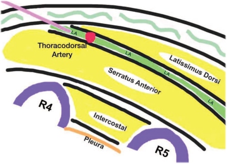
- Superficial thick muscle → Latissimus dorsi
- Deep hyperechoic structure → Serratus anterior muscle
- Deeper bony structures → Ribs
- Pleura (hyperechoic sliding line below ribs)
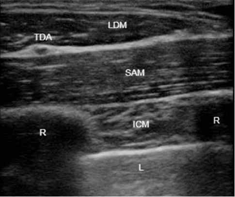
Two techniques
Superficial plane – between LDM and SAM
Deep plane – between SAM and rib
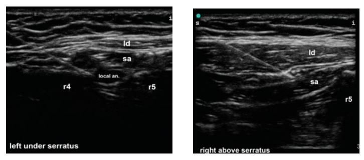
- Using 22 to 25G needle, around 20 to 30 ml of local anaesthetics is given
COMPLICATIONS
- Local anaesthetic systemic toxicity
- Pneumothorax
- Vascular injury
SIGNIFICANCE
- Can be used in management of anterlateral and lateral chest wall pain such as in post herpetic neuralgia, rib fracture
- Analgesia in procedure such as thoracoscopy, VATS
- This patient was proceeded with infiltration of 15ml of 0.5% Ropivacaine and 15ml of 2% Lignocaine with adrenaline in Superficial SAP
- Supplemental systemic analgesics was provided during biospy from parietal pleura
- Parietal pleura is usually spared and hence it was anaethetized using local anaesthetic spray
- Patient remained stable throughout and hence shifted to recovery
- SABP served as an alternative option to GA, minimizing risks of respiratory complications
 Dr Velmurugan
Head of the Department
Department of Anaesthesiology
Kauvery Hospital, Chennai
 Dr Nirmalraj
Final Year – DNB Resident
Department of Anaesthesiology
Kauvery Hospital, Chennai


