Case Details:
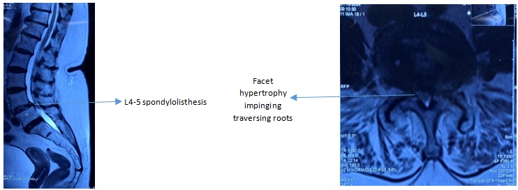
A 79-year-old female presented with radiating pain and paresthesia in both lower limbs, pseudo-claudication pain reduced her walking distance and standing time significantly and had tried medications and epidural steroid injection with little success. Co-morbidities other than obesity include diabetes mellitus, systemic hypertension and hypothyroidism. On examination, she had forward stooping gait, straight leg raise test was negative and MRC grade 4 power in bilateral L5 myotome. Plain radiograph revealed grade I spondylolisthesis at L4-5 lumbar spine with no instability on dynamic stress radiographs. MR imaging(Fig. 1) showed significant facet hypertrophy on both sides encroaching the canal causing bilateral severe foraminal stenosis and moderate – severe central canal stenosis.

Fig. 1: MRI sagittal and axial image
Case Discussion:
Patho-anatomy:
- L4-5 spondylolisthesis – moderate central canal stenosis but no foraminal stenosis
- Facet hypertrophy – severe bony compression of traversing roots (L5) and central canal stenosis
Surgical goal: Decompression of spinal canal and bilateral recess. Since there is no significant back pain and there is no instability in dynamic radiograph, reduction and fusion of spondylolisthesis is not mandatory.
Surgical Options:
- Decompression and fusion: This can be done through conventional open approach as well as minimally invasive approach. Age, obesity and osteoporosis were not in favour of instrumentation and it involves longer hospital stay, blood loss and prolonged recovery period.
- Conventional decompression: it involves detachment of paraspinal muscle attachments and retraction, removal of spinous process and its ligaments, and partial facetal resection on both sides. Although complete decompression can be achieved, this can lead to instability at that motion segment leading to early failure.
- Minimally invasive tubular over the top decompression: tubular retractors are available up-to 11cm length, but the skin to lamina distance is 13-14cm in our patient and hence is not feasible.
- Endoscopic decompression: Stenoscope has the advantage of working length, illumination and magnification. It is least invasive with minimal collateral damage to muscle, ligaments and bone.
Procedure:
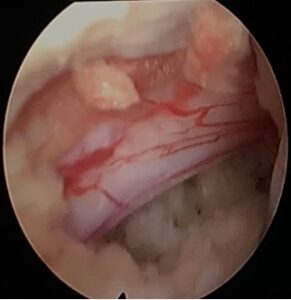
Under general anaesthesia, patient in prone position, endoscopic sleeve docked over right L4/5 facet. Medial 1/3rd of facet burred, lamina (inferior half) of L4 and superior aspect of L5 lamina burred. Base of spinous process burred and over the top decompression done and ligamentum flavum was removed en-bloc. Both lateral recess decompression done with endoscopic burr. Theca and roots found adequately free (Fig. 2).

Fig. 2: Free L5 root after ipsilateral lateral recess decompression
Conclusion:
Full endoscopic uniportal bilateral lumbar decompression is the least invasive procedure for lumbar canal stenosis. It is more valuable in patients with co-morbidities especially obesity as it provides early recovery, minimally blood loss and enables early ambulation.
 Dr. P. Keerthivasan
Consultant Orthopaedic Spine Surgeon
Kauvery Hospital Chennai
 Mr. Balamanimurugan
Physician asst of Dr. Keerthivasan
Kauvery Hospital Chennai
 Dr. Soma Sundar S
Associate Consultant Ortho Spine
Kauvery Hospital Chennai
 Dr. G. Balamurali
Senior Consultant Spine and Neurosurgery
Kauvery Hospital Chennai


