
Diagnostic concordance between cytochemical myeloperoxidase and flow cytometric myeloperoxidase in acute leukemia: A retrospective audit from a tertiary care hospital
Abstract
Background: This study evaluated the diagnostic concordance of in-house cytochemical myeloperoxidase (cMPO) staining against outsourced flow cytometry (fMPO) at Kauvery Hospital, Trichy.
Methods: From an archive of 300 bone marrow cases (January–April 2026), 29 newly diagnosed acute leukemia cases were audited. Manual cMPO results ( ≥ 3% threshold at 100x microscopy) were compared with outsourced immunophenotyping.
Results: The cMPO stain demonstrated 80.0% sensitivity, 100.0% specificity, 100.0% PPV, 90.5% NPV, and an overall accuracy of 93.1%. Complete concordance was achieved in 86.2% (n=25) of cases. Two cMPO-negative AML cases were detected as “dim positive” by flow cytometry. Cohen’s Kappa (κ≈0.84) showed excellent agreement between modalities.
Conclusion: Positive cMPO results offer 100% diagnostic certainty for AML, enabling immediate treatment. However, due to a 20% false-negative rate in poorly differentiated blasts, negative results mandate flow cytometric correlation.
Keywords: Acute leukemia; Acute myeloid leukemia; Myeloperoxidase; Cytochemistry; Flow cytometry; Immunophenotyping.
Introduction
Lineage assignment in acute leukemia is the important step of effective therapeutic planning, prognosis, and risk stratification. Historically, cytochemical Myeloperoxidase (cMPO) staining has been the reliable parameter of hematopathology laboratories for identifying myeloid differentiation, allowing the definitive diagnosis of Acute Myeloid Leukemia (AML).
Based on which the entire field of study was built upon before newer technologies came along.
In the modern diagnostic era, flow cytometric MPO (fMPO) has emerged as a highly sensitive alternative, capable of detecting intracellular MPO proteins at lower thresholds, even in cases with minimal granular maturation (e.g., AML-M0/M1). However, flow cytometry often requires outsourcing in resource-limited or tier-2 city settings, leading to turnaround delays and increased financial burdens on patients.
This study aims to evaluate the diagnostic efficacy and concordance of the in-house cytochemical MPO stain used at Department of Hematopathology, Kauvery Hospital, Trichy, against the outsourced flow cytometric immunophenotyping. The objective is to determine if the traditional glass-slide cytochemical method remains a robust, reliable, and efficient tool for rapid myeloid blast identification in standard clinical workflows.
Materials and Methods
Study Design and Case Selection
A retrospective, archive-based audit was conducted at the Department of Hematopathology, Kauvery Hospital, Trichy. The study sample was derived from a total of 300 bone marrow cases reported over a 4-month period from January 2026 to April 2026. From this archive, 29 newly diagnosed cases of acute leukemia were extracted based on morphological evaluation of bone marrow aspirates.
Cytochemical Staining (cMPO)
In-house cytochemical staining for MPO was performed using standard laboratory protocols (myeloperoxidase staining with diazo dye methods). Stained smears were evaluated under 100x oil immersion microscopy by hematopathologists. Blasts showing ≥ 3% granular cytoplasmic positivity were interpreted as cMPO positive.
Flow Cytometry (fMPO)
Flow cytometric immunophenotyping was outsourced to an outside laboratory. The outsourced flow cytometry reports were collected and archived.
Data Analysis and Statistics
The performance metrics of cMPO were calculated using fMPO as the gold-standard reference. The statistical parameters evaluated included sensitivity, specificity, positive predictive value (PPV), negative predictive value (NPV), overall diagnostic accuracy, and Cohen’s Kappa Coefficient (κ) to measure the strength of agreement between the two modalities. Institutional Ethics Committee approval has been obtained.
Results
Out of the 29 acute leukemia cases evaluated, 8 cases were positive by cytochemical MPO (cMPO) and 21 cases were negative (Table 1).
Table 1: Diagnostic Cross-Tabulation of Manual Cytochemical MPO vs. Flow Cytometric MPO Positivity (n = 29)
| Parameters | fMPO positive | fMPO negative |
|---|---|---|
| cMPO Positive | 8 | 0 |
| cMPO Nega-tive | 2 | 19 |
Table 2: The cross-tabulation with the final flow cytometry diagnosis reveals the following diagnostic distribution:
| cMPO Status | Flow Cytometry Final Diagnosis | fMPO Status | Case Count |
|---|---|---|---|
| Positive (n=8) | Acute Myeloid Leukemia (AML) | Positive | 7 |
| Mixed Phenotype Acute Leukemia (MPAL) | Positive | 1 | |
| Negative (n=21) | B-Acute Lympho-blastic Leukemia (B-ALL) | Negative | 13 |
| T-Acute Lympho-blastic Leukemia (T-ALL) | Negative | 1 | |
| Early T-Cell Pre-cursor ALL (ETP-ALL) | Negative | 1 | |
| Acute Myeloid Leukemia (AML) | Negative | 4 | |
| Acute Myeloid Leukemia (AML) | Dim Pos-itive | 2 |
To evaluate the diagnostic performance of the in-house cytochemical Myeloperoxidase (cMPO) stain against the gold-standard reference of outsourced flow cytometric MPO (fMPO), standard diagnostic accuracy metrics were calculated by cross-classifying the 29 acute leukemia cases. The alignment of the two modalities resulted in 8 True Positive (TP) cases, consisting of 7 cases of Acute Myeloid Leukemia (AML) and 1 case of Mixed Phenotype Acute Leukemia (MPAL) where both testing methods demonstrated positive MPO expressions. The study identified 0 False Positive (FP) cases, as no lymphoid or non-myeloid malignancies exhibited aberrant staining on the cytochemical slides. A total of 19 True Negative (TN) cases were confirmed, encompassing all non-myeloid lineages (13 B-ALL, 1 T-ALL, and 1 ETP-ALL) alongside 4 true MPO-negative AML variants that lacked expression on both testing platforms. Crucially, the analysis revealed 2 False Negative (FN) cases, representing AMLs that were reported as negative under microscopy but were subsequently detected as dim positive by the flow cytometer
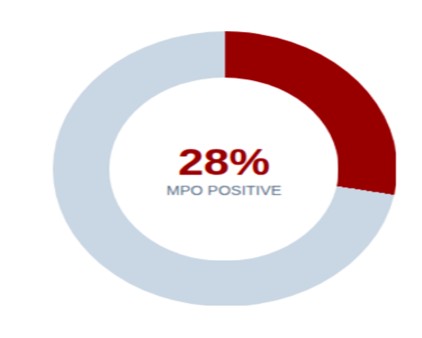
Fig (1): A graphical breakdown of the 29 acute leukemia cases, highlighting a 28% overall MPO positivity rate across both testing platforms in the study cohort.
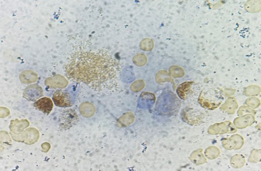
Fig (2): Bone marrow smear under 100x oil-immersion revealing leukemic blasts with moderate-to-strong granular cytoplasmic positivity on manual cytochemical Myeloperoxidase (cMPO) staining.
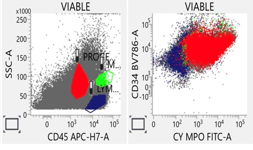
Fig (3): Flow cytometry plot of an AML case showing primary CD45 blast gating alongside moderate-to-bright intracellular MPO and CD34 co-expression, confirming myeloid lineage.
Based on these classifications, the in-house cMPO stain demonstrated a Sensitivity of 80.0%, indicating that while it successfully captures the vast majority of myeloid lineages, it misses hypogranular or poorly differentiated blasts. Conversely, the method achieved a specificity of 100.0% and a Positive Predictive Value (PPV) of 100.0%, confirming that a positive result under the light microscope provides absolute diagnostic certainty for myeloid lineage assignment without the risk of a false alarm. The Negative Predictive Value (NPV) was calculated at 90.5%, reflecting a moderate risk that a negative cytochemical result might mask an underlying dim-positive myeloid leukemia. The Overall Diagnostic Accuracy of the protocol stood at 93.1%, proving that the manual method is highly reliable in a standard clinical workflow. Finally, to evaluate the statistical strength of the agreement between the two diagnostic mediums beyond mere chance, Cohen’s Kappa coefficient was calculated. A calculated Cohen’s Kappa (κ ≈ 0.84) indicates excellent agreement between the in-house cytochemical MPO and flow cytometric MPO analysis.
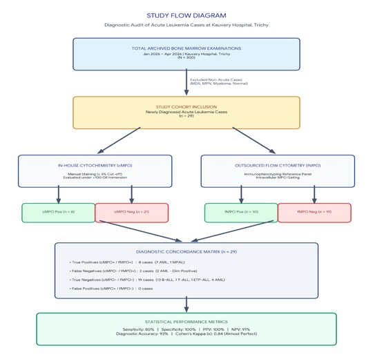
Fig (4): Study Flow diagram.
Discussion
The diagnostic audit at Kauvery Hospital, Trichy, underscores a highly reliable level of clinical utility for traditional, in-house cytochemistry. In our cohort of 29 acute leukemia cases, the manual cytochemical Myeloperoxidase (cMPO) stain achieved an overall diagnostic accuracy of 93%, paired with a flawless specificity and positive predictive value (PPV) of 100%. These metrics demonstrate that when a positive cMPO result is obtained under 100x oil immersion microscopy, the clinical team can definitively diagnose an Acute Myeloid Leukemia (AML) lineage with complete diagnostic certainty. Our observed overall concordance rate of 93% stands out as superior to the 86% overall concordance rate documented by Belurkar et al. [1]. Complete concordance was achieved in 86% of our cases (n = 25), reflecting cases like Acute Lymphoblastic Leukemia (ALL) and standard AML where both platforms fully agreed, heavily surpassing the 58% complete concordance noted by their group [1]. Partial concordance—where both platforms recognized an AML lineage but differed strictly on staining intensity—was seen in 7% (n = 2). Non-concordance was restricted to just 7% (n = 2), matching closely with Belurkar’s low 4% non-concordance threshold [1]. The critical operational limitation of cytochemistry highlighted by our audit is its sub-optimal sensitivity (80%), driven by 2 false-negative manual readings. This specific pitfall was analyzed at length in a comparative study conducted at the All-India Institute of Medical Sciences (AIIMS), New Delhi, where Jain et al. [4] highlighted that significant MPO discrepancies frequently occur across testing platforms due to variable analytical sensitivities. Flow cytometry easily catches the dim positives missed on glass slides because it utilizes laser-based fluorochromes to identify low-density intracellular MPO proteins [4]. Our findings are also closely echoed by Goud et al. [3] in Cureus (2024), who reported that combined morphological and cytochemical analysis successfully rendered accurate diagnoses in 93% of acute leukemia cases. Similar to our 2 discordant cases that were missed by microscopy due to a dim positive flow cytometric MPO (fMPO) profile, the Cureus investigators observed that their manual diagnostic failures occurred exclusively within poorly differentiated sub-types, specifically M0 and M1-AML variants [3]. From a clinical standpoint, given the 100% specificity and PPV observed in our results, an initial positive cMPO slide allows the medical team to initiate induction therapy for AML without waiting for outsourced immunophenotyping reports, thereby drastically reducing turnaround time for critically ill patients. Furthermore, despite the growing popularity of flow cytometry, the modality remains hindered by economic realities like costly outsourcing fees, specialized infrastructure needs, turnaround delays, and technical limitations involving clotted samples or dry-tap aspirates where cytochemistry thrives [4]. A well-standardized, in-house cMPO protocol functions as a powerful, near-flawless screening gatekeeper that protects resource-limited setups from unnecessary diagnostic delays. Finally, although the current WHO 2022 Classification [7] and the International Consensus Classification (ICC) [6] place an increasingly strict reliance on quantitative immunophenotyping for complex lineages, a definitive consensus percentage cut-off for MPO staining intensity in flow cytometry remains to be established.
Limitations
- Single-Center Study: The audit was conducted exclusively as a single-center retrospective study.
- Small Sample Size: The study cohort was limited to a small sample size (n = 29).
- Outsourced Testing: Flow cytometry was performed at an external reference laboratory rather than in-house.
- No Outcome Data: The study did not include long-term clinical outcome or patient survival analysis.
Conclusion
This institutional audit confirms that the in-house cytochemical MPO stain used at Kauvery Medical Centre, Trichy, is a highly efficient, accurate (93.1%), and cost-effective methodology for the initial diagnosis of acute myeloid leukemia. Given its 100% positive predictive value, a positive cMPO stain under 100x microscopy acts as a definitive diagnostic tool for calling a blast a myeloid blast, mitigating turnaround delays associated with outsourced reference laboratories.
We conclude that, in-house cytochemical MPO remains a highly specific, rapid and cost-effective screening tool for AML in resource-limited settings. However, negative or equivocal cytochemical findings should always be confirmed using flow cytometric immunophenotyping [1, 3, 4]
References
- Belurkar S, Mantravadi H, Manohar C, Kurien A. Correlation of morphologic and cytochemical diagnosis with flowcytometric analysis in acute leukemia. J Cancer Res Ther. 2013;9(1):71-79. doi:10.4103/0973-1482.110378.
- Putti MC, Basso G, Rondelli R, Aric M, Biondi A, Cordero di Montezemolo L, et al. Cell lineage classification derived from flow cytometry and cytochemical stains in childhood acute leukemia. Med Pediatr Oncol. 2001;36(2):265-271. doi:10.1097/00043426-200102000-00004.
- Goud RE, Kumar S, Patel A, Sharma M, Verma N. Diagnosis and correlation of acute leukemia by morphology, cytochemistry, and flow cytometry immunophenotyping. Cureus. 2024;16(2):e54126. doi:10.7759/cureus.54126.
- Jain S, Malik F, Sharma S, Singh T. Myeloperoxidase detection by cytochemistry, immunohistochemistry and flow cytometry in acute myeloid leukemia: A comparative study from a tertiary care center. Indian J Hematol Blood Transfus. 2017;33(4):461-468. doi:10.1007/s12288-017-0849-1.
- Vardiman JW, Thiele J, Arber DA, Brunning RD, Borowitz MJ, Porwit A, et al. The 2008 revision of the World Health Organization (WHO) classification of myeloid neoplasms and acute leukemia: rationale and important changes. Blood. 2009;114(5):937-951. doi:10.1182/blood-2009-03-209262.
- Khoury JD, Solary E, Abla O, Akkari Y, Alaggio R, Apperley JF, et al. The International Consensus Classification of Myeloid Neoplasms and Acute Leukemias: integrating morphologic, clinical, and genomic data. Blood. 2022;140(11):1200-1228. doi:10.1182/blood.2022015857.
- Khoury JD, Solary E, Abla O, Akkari Y, Alaggio R, Apperley JF, et al. The 5th edition of the World Health Organization Classification of Haematolymphoid Tumours: Myeloid and Histiocytic/Dendritic Neoplasms. Leukemia. 2022;36(7):1703-1719. doi:10.1038/s41375-022-01613-1.
- Manivannan P, Puri V, Somasundaram V, Purohit A, Sharma RK, Dabas M, et al. Can threshold for MPO by flow cytometry be reduced in classifying acute leukaemia? A comparison of flow cytometric and cytochemical myeloperoxidase using different flow cytometric cut-offs. Hematology. 2015;20(8):455-461. doi:10.1179/1607845414Y.0000000223.
- Swerdlow SH, Campo E, Harris NL, Jaffe ES, Pileri SA, Stein H, et al., editors. WHO Classification of Tumours of Haematopoietic and Lymphoid Tissues. 4th ed. Lyon: International Agency for Research on Cancer; 2008.
- Bennett JM, Catovsky D, Daniel MT, Flandrin G, Galton DA, Gralnick HR, et al. Proposals for the classification of the acute leukaemias. French American-British (FAB) co-operative group. Br J Haematol 1976;33(4):451-458. doi:10.1111/j.1365-2141.1976.tb03563