
Tuberculous pericarditis presenting with cardiac tamponade and refractory shock in an immunocompetent adult: A case report and review of diagnostic challenges
Abstract
Background: Tuberculous pericarditis is an uncommon yet potentially fatal manifestation of extrapulmonary tuberculosis, accounting for approximately 1–2% of all tuberculosis cases. Delayed diagnosis frequently results in life-threatening complications including cardiac tamponade, constrictive pericarditis, and multiorgan failure. Establishing microbiological confirmation remains difficult because of the paucibacillary nature of the disease and the limited sensitivity of conventional diagnostic techniques.
Case Presentation: We report the case of a 58-year-old man with type 2 diabetes mellitus, chronic kidney disease, systemic hypertension, and ischemic heart disease who presented with a two-month history of fever, chronic cough, weight loss, and progressive pericardial effusion. Emergency pericardiocentesis yielded 200 mL of straw-coloured pericardial fluid. Laboratory analysis demonstrated an adenosine deaminase (ADA) level of 71 U/L and acid-fast bacilli positivity. Despite emergency drainage, thoracotomy, intensive care support, and resuscitative measures, the patient developed refractory shock and expired. Owing to rapid clinical deterioration, definitive antitubercular therapy could not be initiated before the patient developed refractory shock.
Conclusion: This case highlights the importance of maintaining a high index of suspicion for tuberculous pericarditis in tuberculosis-endemic regions and underscores the indispensable role of the clinical microbiology laboratory in facilitating early diagnosis through rapid microbiological investigations.
Keywords: Tuberculous pericarditis; Cardiac tamponade; Extrapulmonary tuberculosis; Pericardial fluid; ADA; Acid-fast bacilli; Septic shock
Introduction
Tuberculosis remains a major public health problem worldwide, with India accounting for nearly one-fourth of the global disease burden. Although pulmonary disease predominates, extrapulmonary tuberculosis contributes significantly to morbidity and mortality.
Pericardial involvement is relatively uncommon but carries considerable clinical significance because delayed diagnosis may result in cardiac tamponade, constrictive pericarditis, septic shock, and death. TB pericarditis constitutes approximately 1–4% of acute pericarditis in endemic countries. Mortality reaches 40–60% without treatment.
Tuberculous pericarditis typically results from contiguous spread from mediastinal lymph nodes, hematogenous dissemination, or direct extension from pulmonary lesions. The disease is usually paucibacillary, making microbiological confirmation difficult. Consequently, diagnosis often relies upon a combination of clinical suspicion, imaging, biochemical markers such as ADA, molecular methods, mycobacterial culture, and histopathology.
We report a fatal case of tuberculous pericarditis presenting with cardiac tamponade and refractory shock to emphasize the diagnostic challenges encountered by clinicians and microbiologists.
Case Presentation
A 58-year-old gentleman presented with intermittent fever, chronic cough for two months, evening rise of temperature, anorexia, and significant weight loss. Two months before presentation, the patient had been admitted elsewhere for multidrug-resistant Escherichia coli urinary tract infection and was subsequently evaluated for moderate pericardial effusion. Pericardiocentesis had been advised but was initially declined.
Past medical history included:
- Type 2 Diabetes Mellitus
- Chronic Kidney Disease
- Systemic Hypertension
- Triple Vessel Coronary Artery Disease
- Percutaneous coronary intervention involving the LAD and LCX in 2017
At presentation to our institution, he was hypotensive and admitted to the coronary intensive care unit for further evaluation.
Clinical Examination
The patient was conscious and oriented.
Vital signs demonstrated:
- Blood pressure: 60 mmHg systolic/? diastolic
- Pulse rate: 90 beats/minute
- Respiratory rate: 19/minute
- Oxygen saturation: 98% on room air
- Local examination: No pedal edema
- Cardiovascular examination: Muffled heart sounds without clinical evidence of overt heart failure.
- Respiratory system: Normal vesicular breath sounds
- P/A soft. No hepatomegaly.
Investigations
Baseline laboratory investigations done.
- 2 Sputum AFB samples negative
- Elevated CRP, ESR,
- Elevated SGOT, SGPT, lactate
- Hb 11.3g/dl
- Creatinine 2.85mg/dl
- Urea 124mg/dl
- NT Pro BNP (N – Terminal B Type) 8630pg/ml
Echocardiography revealed:
- Moderate circumferential pericardial effusion
- Preserved left ventricular function
- Echocardiographic features suggestive of cardiac tamponade
Emergency pericardiocentesis was performed. Approximately 200 mL of straw-coloured pericardial fluid was aspirated.
Pericardial fluid analysis demonstrated:
| Parameter | Result |
|---|---|
| Appearance | Straw coloured |
| ADA | 71 U/L |
| WBC Count | 4720 cells/mm³ |
| Neutrophils | 71.6% |
| Other Mononuclear cells | 28.4% |
| Light’s criteria | Suggestive of transudative pericardial effusion. |
| AFB smear | Positive |
The elevated ADA together with AFB positivity strongly supported the diagnosis of tuberculous pericarditis.
Cytology Report: Smears studied are cellular showing predominantly sheets of neutrophils, admixed with neutrophilic debris, cyst macrophages, histiocytes and occasional scattered medium sized mononuclear cells exhibiting mild to moderate anisonucleosis with hyperchromatic nuclei and eosinophilic cytoplasm in a dirty and haemorrhagic background. Atypia of Undetermined significance (AUS)with dense inflammation and haemorrhage. Advised clinico-radiological correlation
Gram Stain – Few pus cells, no organisms seen
Culture Report: No growth after 48hrs of aerobic incubation
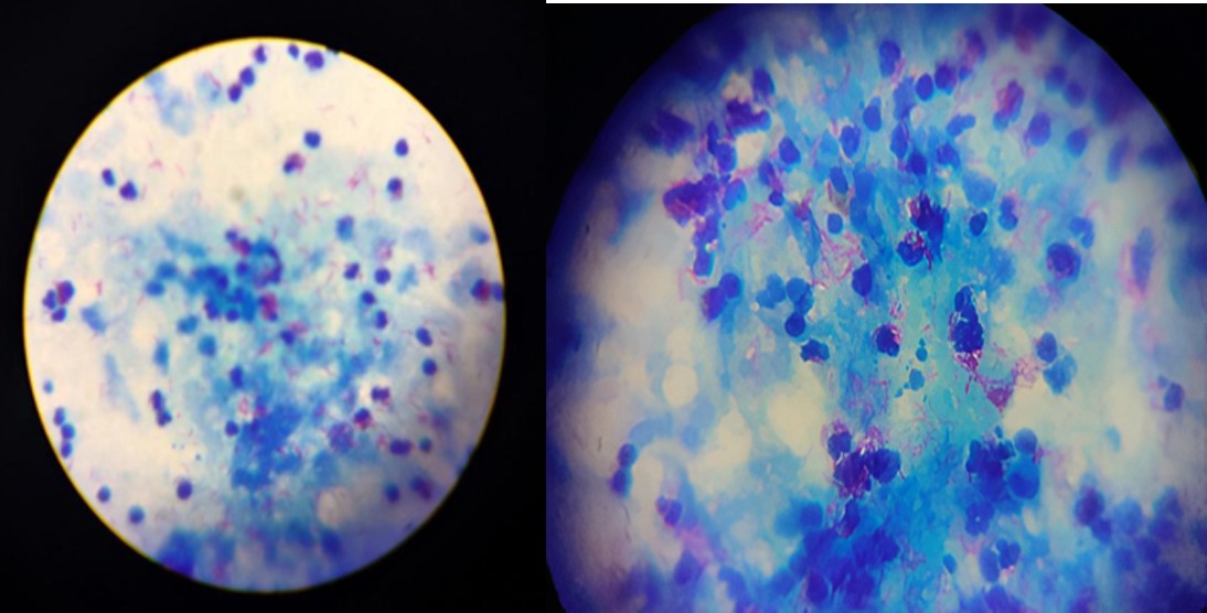
Fig (1): Acid fast stain of pericardial fluid showing Acid fast Bacilli
Treatment given
The patient underwent emergency pericardiocentesis using the subxiphoid approach. Despite drainage, echocardiography demonstrated persistent thick organized collections not amenable to catheter drainage. Cardiothoracic surgical consultation was obtained, and emergency thoracotomy was performed. The patient received vasopressor support, intensive care monitoring, and advanced cardiac life support. Unfortunately, refractory shock, severe metabolic acidosis, and cardiac arrest ensued despite aggressive management. Owing to rapid clinical deterioration, definitive antitubercular therapy could not be initiated before the patient developed refractory shock.
Outcome and Follow-up
The patient developed progressive multiorgan dysfunction culminating in bradycardia, asystole, and death despite maximal supportive care.
Differential Diagnosis: The differential diagnoses included:
- Viral pericarditis
- Pyogenic bacterial pericarditis
- Malignant pericardial effusion
- Autoimmune pericarditis
- Uremic pericarditis
- Tuberculous pericarditis
The constitutional symptoms together with elevated ADA and AFB positivity established tuberculosis as the most likely diagnosis.
Discussion
Tuberculous (TB) pericarditis occurs when Mycobacterium tuberculosis infects the pericardial sac. It presents insidiously with fatigue, night sweats, fever, and chest pain. Complications include cardiac tamponade and constrictive pericarditis. Standard management requires 6 months of antitubercular therapy (RIPE) and sometimes adjunctive corticosteroids
Clinical Stages
Tuberculous pericarditis generally progresses through three to four stages, though patients may present at any point:
Dry Stage: The initial phase, characterized by fibrinous exudation and pericardial friction rubs; pericardial effusion is absent.
Effusive Stage: Fluid builds up (pericardial effusion), frequently resulting in a “water bottle” heart shadow on a chest X-ray. If fluid accumulates rapidly or in large volumes, it can lead to cardiac tamponade, compressing the heart.
Effusive-Constrictive Stage: A transitional phase where the heart is compressed both by fluid and by the thickening and inflammation of the pericardium.
Constrictive Stage: The fluid is reabsorbed, but the pericardium becomes heavily scarred, rigid, and sometimes calcified. This hard casing prevents the heart from stretching and filling properly, leading to heart failure symptoms like leg swelling and ascites.
Diagnosis
Diagnosis can be challenging due to non-specific symptoms. Key diagnostic steps include:
Imaging: Echocardiography and chest CT scans are critical to visualize pericardial thickening, fluid accumulation, or constriction.
Pericardiocentesis: Extracting and analyzing the pericardial fluid. Fluid analysis typically reveals a lymphocytic exudate with elevated protein and high adenosine deaminase (ADA) activity.
Microbiological Discussion: Pericardial tuberculosis represents one of the most difficult forms of extrapulmonary tuberculosis to diagnose microbiologically.
Acid-fast stain: Sensitivity generally ranges from 10–40%. Positive smears, as observed in the present case, strongly support the diagnosis but occur infrequently because pericardial fluid usually contains very low bacillary loads.
GeneXpert MTB/RIF: Although not performed in this patient, GeneXpert has become the preferred rapid diagnostic modality owing to its high specificity (>95%) and ability to detect rifampicin resistance.
Confirmation: The definitive diagnosis requires identifying tubercle bacilli via acid-fast bacilli (AFB) smear, culture, or PCR of the pericardial fluid or tissue biopsy.
Tuberculous pericarditis remains associated with mortality rates approaching 40% despite contemporary management.
The diagnosis is frequently delayed because:
- Constitutional symptoms are nonspecific.
- Pericardial fluid contains few organisms.
- Smear microscopy has poor sensitivity.
- Culture requires several weeks.
- Clinical suspicion is often low.
The present patient exhibited several classic features suggestive of tuberculosis, including prolonged fever, chronic cough, weight loss, elevated ADA, and AFB-positive pericardial fluid.
The elevated ADA level (71 U/L) substantially exceeded the commonly accepted diagnostic threshold of 40 U/L reported in multiple meta-analyses.
Early communication between microbiology laboratories and clinicians is critical because prompt microbiological confirmation facilitates early initiation of antitubercular therapy and may improve survival.
Treatment and Management
Treatment primarily focuses on clearing the infection and preventing or relieving heart failure:
Antitubercular Therapy (ATT): The standard regimen mirrors pulmonary TB: a 2-month intensive phase with rifampin, isoniazid, pyrazinamide, and ethambutol, followed by a 4-month continuation phase with rifampin and isoniazid.
Corticosteroids: The use of adjunctive corticosteroids (such as prednisone) is highly debated but often recommended for patients to reduce inflammation, minimize fluid recurrence, and potentially lower the risk of developing constrictive pericarditis. [1, 2, 3]
Surgical Intervention: If patients develop life-threatening cardiac tamponade or persistent constrictive pericarditis, surgical drainage or a partial/total pericardiectomy (removal of the thickened pericardium) is required.
Learning Points
- Tuberculosis should always be considered in patients with unexplained pericardial effusion in endemic regions.
- ADA remains an important adjunctive diagnostic biomarker.
- GeneXpert should be performed routinely on pericardial fluid whenever feasible.
- Mycobacterial culture should always accompany molecular testing.
- Persistent, organized collections frequently require surgical intervention.
- Close collaboration between cardiology, cardiothoracic surgery, pathology, and microbiology is essential.
Patient Perspective: The patient initially declined pericardiocentesis during an earlier hospitalization. This case emphasizes the importance of counselling patients regarding the potential consequences of delaying invasive diagnostic procedures when clinically indicated.
Informed Consent: Written informed consent for publication should be obtained from the patient’s next of kin prior to manuscript submission.
Conflict of Interest: The authors declare no conflict of interest.
Funding: None.
Acknowledgement
- Thilagavathy MD, (Microbiology): Conceptualization, Writing – original draft, review & editing.
- Rajendran M: Proving Clinical information’s
- Kabilan SJ: Providing Clinical information’s
- Thialagavathi MSc: Laboratory procedures and Technical support
References
- Mayosi BM, Ntsekhe M, Bosch J, et al. Prednisolone and Mycobacterium indicus pranii in Tuberculous Pericarditis. N Engl J Med. 2014;371:1121–1130.
- Adler Y, Charron P, Imazio M, et al. 2015 ESC Guidelines for the diagnosis and management of pericardial diseases. Eur Heart J. 2015;36:2921–2964.
- World Health Organization. Consolidated Guidelines on Tuberculosis. Geneva: WHO; 2024.
- Reuter H, Burgess LJ, Doubell AF. Tuberculous pericarditis. J Am Coll Cardiol. 2006;48:133–141.
- Sharma SK, Mohan A. Extrapulmonary tuberculosis. Indian J Med Res. 2004;120:316–353.
- Golden MP, Vikram HR. Extrapulmonary tuberculosis: An overview. Am Fam Physician. 2005;72:1761–1768.
- Fowler NO. Tuberculous pericarditis. JAMA. 1991;266:99–103.