
When the masquerader drops his mask: A case of neurocysticercosis
Abstract
Background: Neurocysticercosis is an infection of the central nervous system affecting the brain and the meninges. Although, this infection can be treated medically, more often this condition presents as a seizure disorder. Neurocysticercosis is caused by infestation by Taenia solium, commonly known as pork tapeworm.
Case Presentation: A 27-year-old male patient presented with recent onset seizures and giddiness. Investigations revealed a cystic lesion with calcification in right inferior temporal gyrus region suggestive of a glioneuronal neoplasm on imaging. The histopathological examination revealed the presence of a non-viable cysticercus larva confirming Neurocysticercosis.
Conclusion: This case highlights a rare presentation of neurocysticercosis mimicking a glioneuronal neoplasm on imaging, emphasizing the importance of histopathology as the gold standard for diagnosis.
Keywords: Neurocysticercosis, seizure disorder
Introduction
Neurocysticercosis is an infection caused by the larval worm of Taenia solium. This is the most common helminthic infection of the central nervous system affecting the brain and meninges. The infection usually remains asymptomatic for a long period before presenting as sudden onset of seizures. Neuro imaging remains the main diagnostic modality for neurocysticercosis. The disease presents as intraparenchymal, extraparenchymal and ocular forms; of which intraparenchymal presentation is the most common.
This disease is common in India, Nepal, sub-Saharan Africa and Latin America. Humans are the only
definitive host for the parasite. Humans and pigs act as intermediate hosts. Ingestion of undercooked pork meat containing cysticerci causes intestinal Taeniasis, whereas human cysticercosis results from ingestion of Taenia solium eggs by faecal-oral transmission[1].
Neuroimaging Criteria
Major criteria
- Cystic lesion(s) with no discernible scolex
- Enhancing lesions
- Multilobulated subarachnoid cystic lesions
- Solid calcified lesion (usually < 1 cm)
Minor criteria
- Obstructive hydrocephalus
- Abnormal enhancement of basal leptomeninges
Clinical/Epidemiological Criteria
Major criteria
- Serologic evidence of specific anticysticercal antibodies or cysticercal antigens by standardized immunodiagnostic tests
- Extra neural cysticercosis
- Household contact with taeniasis
Minor criteria
- Suggestive clinical manifestations
- Residence in an endemic area
- Definite neurocysticercosis diagnosis is based on neuroimaging and clinical/epidemiologic criteria2
- One absolute criterion or
- Two major neuroimaging criteria and epidemiological exposure criteria or
- One major imaging criterion and two epidemiological exposure criteria and exclusion of diseases with a similar radiologic presentation
Case Presentation
A 27-year-old male patient presented with history of recent onset seizures, jerky movements of limbs with loss of consciousness for few minutes. There was history of giddiness on and off since a week. There was no history of seizures in the past. There was no history of exposure or travel to endemic areas. On examination his vital signs were within normal limits. CNS examination showed a GCS 15/15 and no focal neurologic deficit. Respiratory and cardiovascular system examination was within normal limits.
MRI brain plain and contrast and MRS were done and showed focal small relatively well circumscribed cystic lesion with mural T2 hypointense component and eccentric calcifications in the right third inferior temporal gyrus region with minimal perilesional vasogenic oedema. Eccentric thin peripheral enhancement with enhancing mural/septae were noted. These features were suggestive of low-grade glioneuronal tumour like dysembryoplastic neuroepithelial tumour /ganglioglioma. Sleep deprived EEG was done showed sleep stage II with superimposed fronto-temporal sharp and discharges. Necessary blood investigations were done and were within normal limits.[2]
Since the imaging findings were in favour of a glioneuronal neoplasm, he was advised surgical treatment. After obtaining informed consent, he underwent right parietotemporal craniotomy with total excision of space occupying lesion under neuronavigation and electrocorticography guided surgery. The surgeon requested a frozen section examination for intra-operative consultation. However, in view of presence of calcifications, the frozen section examination was deferred and proceeded for permanent sections. The postoperative course of this patient was uneventful.
On histopathological examination, sections showed glial tissue with a cystic cavity containing a non-viable cysticercus with a hyalinised wall having a tegument with finger like projections and microvilli on the surface. Multiple stromal calcareous bodies were seen subjacent to the tegument. There was surrounding granulomatous reaction composed of lymphocytes, plasma cells, histiocytes including epithelioid histiocytes, plenty of eosinophils and few neutrophils. The surrounding brain parenchyma showed reactive gliosis.
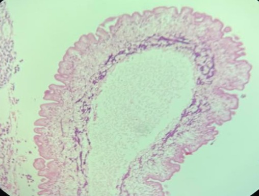
Fig (1): H&E ×100 magnification, non-viable cysticercus with a hyalinised wall having a tegument with finger like projections and microvilli on the surface
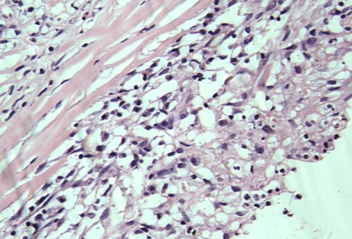
Fig (2): H&E ×400 magnification showing surrounding granulomatous reaction composed of lymphocytes, plasma cells, histiocytes including epithelioid histiocytes, numerous eosinophils and few neutrophils.
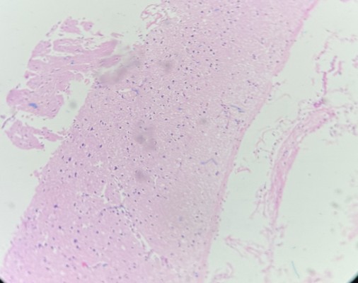
Fig (3): H&E ×100 magnification showing surrounding brain tissue showing reactive gliosis.
Discussion
Cysticercosis has an extremely variable presentation, with symptoms dependent on the tissues that Taenia solium has infected, with neurocysticercosis being diagnosed when tissues in the central nervous system are affected3. This variability in symptomatology extends to the severity of these symptoms as well, ranging from completely asymptomatic to acutely life-threatening. Most common mode of presentation of neurocysticercosis is as seizure disorder. However, the other forms of the disease can present as increased intracranial tension or cognitive decline. An important clue to diagnosis is the presence of history of travel to endemic areas. In our case, the imaging was suggestive of glioneuronal tumour because of the presence of cystic areas with focal calcification against the absolute neuroimaging criteria for neurocysticercosis. Similar imaging features can be seen in other conditions such as tuberculoma, mycotic infections and low-grade glial neoplasms. Since there was no history of previous exposure or travel to endemic areas and atypical presentation of the infection as a cystic lesion with calcification, histopathological examination helped in conclusive diagnosis. Glioneuronal tumours tend to present in supratentorial area, usually in the frontal and temporal lobes, and are closely related to the ventricles. Imaging features include mixed cystic and solid mass, cystic mass with mural nodule, pure cystic mass, and pure solid mass[5].
Treatment with antihelminthic drugs like albendazole and praziquantel has been recommended. These medications may kill the larva but elicit an inflammatory response. Hence concurrent anti-inflammatory therapy is also initiated.
Conclusion
This case is presented because of the unusual radiological appearance of the lesion, which closely resembled a low-grade glioneuronal neoplasm and therefore created a diagnostic dilemma. Such imaging characteristics can make differentiation between neoplastic and non-neoplastic lesions challenging, particularly when the lesion has a relatively slow-growing or benign-appearing appearance. Awareness of these uncommon radiological presentations is important to prevent diagnostic delay and to ensure that appropriate investigations and management are undertaken at the earliest opportunity. Prompt recognition and accurate diagnosis may contribute to better clinical outcomes by allowing timely treatment and reducing the risk of persistent or progressive neurological dysfunction. In addition, the anatomical site of the lesion is an important determinant of the patient’s clinical presentation, as involvement of different regions can result in distinct neurological symptoms and signs. The location may also influence the feasibility and choice of treatment, the potential for postoperative morbidity, and ultimately the patient’s prognosis. Through this case, we aim to highlight the diagnostic challenges associated with this uncommon imaging presentation and emphasize the importance of integrating radiological findings with clinical and pathological information when evaluating lesions that mimic low-grade glioneuronal tumors.
References
- Neurocysticercosis: A review of current status and management; Sumit Sinha , B.S. Sharma, Journal of clinical neuroscience, Volume 16, Issue 7p867-876July 2009
- Clin Microbiol Rev 2020;33:e00085, Acta Neurol Scand 1997;96:76, J Neurol Sci 2017;372:202
- Del Brutto OH, Neurocysticercosis: Neurohospitalist, 2014; 4; 205-12
- Del Brutto OH, García HH, Taenia solium cysticercosis – the lessons of history: J Neurol Sci, 2015; 359; 392-95
- Park CK, Phi JH, Park SH (2015) Glial tumors with neuronal differentiation. Neurosurg Clin N Am 26(1):117–138