
Mammary myofibroblastoma mimicking malignancy: A case report and diagnostic approach
Abstract
Background: Mammary myofibroblastoma (MFB) is a rare, benign mesenchymal tumor of the breast predominantly affecting older men and postmenopausal women. Due to overlapping clinical, radiological, and core needle biopsy findings, it frequently simulates malignant breast lesions, posing significant diagnostic challenges. We present a case of a 59-year-old female with a radiologically suspicious lesion. Final histopathological examination demonstrated a vaguely lobulated proliferation of bland spindle cells set in a densely collagenized stroma. Immunohistochemistry performed confirmed the diagnosis of mammary myofibroblastoma. In conclusion, MFB is a rare benign stromal tumor that can clinically and radiologically mimic breast carcinoma. Surgical excision remains curative, and a targeted immunohistochemical panel is essential to differentiate MFB from other spindle cell lesions of the breast.
Keywords: Mammary myofibroblastoma; Spindle cell lesion; Breast mesenchymal neoplasm
Introduction
Mammary myofibroblastoma (MFB) is a distinct, rare benign stromal tumor of the breast originally described by Wargotz et al. in 1987[1]. Unlike most breast malignancies and common benign tumors such as fibroadenomas, MFB shows a striking predilection for older men and postmenopausal women [2, 3]. Clinically and radiologically, MFB often presents as a well-circumscribed, mobile mass and may mimic malignant lesions or other stromal neoplasms.
Histologically, MFB is characterized by a proliferation of bland spindle cells within a collagenous stroma containing varying amounts of adipose tissue [4, 5]. Accurate diagnosis relies heavily on characteristic immunohistochemical profiles, notably diffuse positivity for CD34 and desmin, which help rule out malignant mimickers such as metaplastic carcinoma and fibromatosis [4, 6]. We report a classic case of mammary myofibroblastoma in a 59-year-old female, highlighting its clinical presentation, histopathological features, and the diagnostic utility of immunohistochemistry in differentiating it from other spindle cell lesions of the breast.
Case Presentation
A 59-year-old female presented with complaints of a left breast lump associated with mild pain for the past six months. There was no history of rapid increase in size, nipple discharge, skin changes, or constitutional symptoms.
On local examination, a fairly well defined, firm lump measuring 2×2 cm was palpable in the lower outer quadrant of the left breast. The overlying skin was normal, and no palpable axillary lymphadenopathy was detected. General physical examination and systemic evaluation were within normal limits.
Radiological evaluation classified the lesion as a BI-RADS 3 (probably benign), raising suspicion of malignancy. A subsequent tru-cut needle biopsy was performed which showed ductal hyperplasia with foci of sclerosing adenosis. In view of the clinical and radiological findings, a wide local excision of the lump was planned and performed.
Pathological Findings
- Gross Examination: The surgical specimen revealed a well-circumscribed, unencapsulated lesion measuring 2.3×1.5×1.5 cm. The cut surface was whitish to yellowish, lobular, and demonstrated dense fibrous areas. No hard areas, hemorrhage, or necrosis were identified. All surgical margins were grossly free of tumor.
- Microscopy: Histological examination showed a vaguely lobulated lesion composed of intersecting fascicles of bland spindle cells. The cells have scant to moderate cytoplasm, relatively uniform nuclei with vesicular chromatin, and inconspicuous nucleoli. These cellular areas were set within an abundant, densely collagenized stroma interspersed with mast cells, chronic inflammatory cells, and small blood vessels. Focal areas of edema, entrapped adipose tissue, and a few benign duct-lobular units were noted. The periphery showed benign breast parenchyma with mild adenosis and ductal hyperplasia. Mitotic figures were 2/10hpf, and all microscopic margins were clear. (Fig 1)
- Immunohistochemistry (IHC): To evaluate the lesion and distinguish it from relevant spindle-cell and epithelial mimickers, an immunohistochemical panel was performed, with the following results (Fig 2):
- CD34: Diffusely positive in tumor cells.
- Desmin: Diffusely positive in tumor cells.
- Smooth Muscle Actin (SMA): Non-specific nuclear staining.
- Ki-67 Proliferation Index: Very low, less than 3%.
- Pancytokeratin and p63: Negative, arguing against epithelial differentiation and metaplastic carcinoma.
- β-catenin: Negative with no nuclear staining observed, arguing against mammary-type fibromatosis.
Based on these morphological and immunohistochemical findings, a diagnosis of mammary myofibroblastoma was established. The postoperative recovery was uneventful.
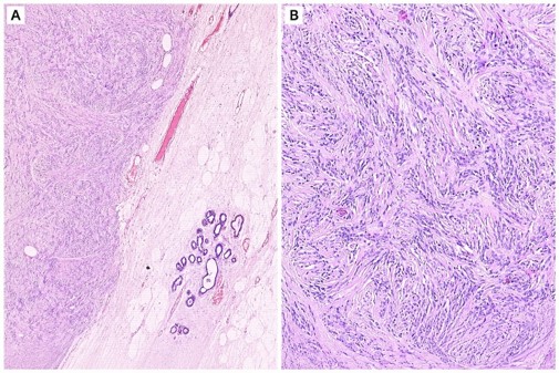
Fig (1): A. shows benign breast tissue with a vaguely lobulated, fairly circumscribed spindle-cell lesion. H&E ×40. B. The image shows vague fascicles and haphazardly arranged sheets of bland spindle cells set in a background of densely abundant collagenized stroma. H&E ×100.
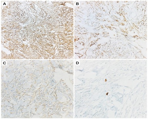
Fig (2): A. CD34 immunohistochemical stain highlights the spindle cells diffusely and strongly. IHC ×100. B. Desmin immunohistochemical stain highlights the spindle cells almost diffusely. IHC ×100. C. Beta-catenin immunohistochemical stain shows cytoplasmic positivity. IHC ×100. D. Ki-67 immunohistochemical stain highlights very few cells (<3%). IHC ×100.
Discussion
Mammary myofibroblastoma is an uncommon benign stromal neoplasm of the breast [2]. While it classically occurs in older men (with a male-to-female ratio of approximately 8:1), postmenopausal women are also affected, as demonstrated in our patient [2, 3]. Because MFB often presents as a firm, circumscribed nodule, it frequently mimics fibroadenoma, phyllodes tumor, or carcinoma both clinically and radiologically [3]. In this case, the lesion was categorized as BI-RADS 3, emphasizing the diagnostic challenge it presents on imaging
The histological spectrum of MFB can be varied, often displaying classic, cellular, infiltrative, epithelioid, or collagenized variants [2]. The classic variant, as observed in our case, features bland spindle cells arranged in fascicles within a collagenous stroma interspersed with adipose tissue [1, 4, 5]. The presence of admixed adipose tissue is an important diagnostic clue, though not universally present
Because spindle cell lesions of the breast may show considerable histologic overlap, immunohistochemistry is an important adjunct to morphological assessment in establishing the diagnosis. MFBs characteristically show strong and diffuse expression of CD34 and desmin, while typically lacking cytokeratins and p63, which reliably excludes metaplastic carcinoma [4, 6]. Furthermore, negative nuclear β-catenin staining differentiates MFB from mammary fibromatosis, which exhibits nuclear accumulation. The extremely low proliferation index (Ki-67 <3%) and sparse mitoses in this case further support its benign nature.
Although cytogenetic abnormalities have been described in mammary myofibroblastoma and its variants [5], the present case showed typical morphological and immunohistochemical features; therefore, cytogenetic studies were not performed.
Complete surgical excision is generally curative, and recurrence following adequate excision is rare [2, 3].
Conclusion
Mammary myofibroblassstoma is a rare benign mesenchymal tumor that can closely simulate breast malignancy on clinical and radiological evaluation [2, 3]. A high index of suspicion, thorough histopathological examination, and an appropriate immunohistochemical panel—including CD34 and desmin positivity, together with negative epithelial markers and absence of nuclear β-catenin staining are important for distinguishing mammary myofibroblastoma from its spindle-cell mimickers and guiding appropriate surgical management [4, 6].
References
- Wargotz ES, Norris HJ, Sesterhenn IA. Fibroblastoma and myofibroblastoma of the breast. Am J Surg Pathol. 1987;11(1):38-46.
- Magro G, Fraggetta F, Torrisi A, Cavallaro A, Righi R, Parenti R. Mammary myofibroblastoma: a report of 7 cases with a comprehensive review of the literature. 2000;108(11):752-60.
- Chiarelli M, Di Mauro S, Gerardi C, et al. Myofibroblastoma of the breast: a case report and review of the literature. Int J Surg Case Rep. 2017;37:168-71.
- McMenamin ME, Fletcher CDM. Mammary myofibroblastoma: an stromal lesion with a putative myofibroblastic differentiation. Am J Surg Pathol. 2001;25(8):1022-9.
- Siziopikou KP, Prioleau J, Okoroh J, Schnitt SJ. Mammary myofibroblastoma: report of a case with a complex karyotype and review of the literature. Mod Pathol. 1998;11(11):1142-6.
- Hasegawa T, Yokoyama R, Matsuno Y, Shimoda T, Hirohashi S. Myofibroblastoma of the breast: an immunohistochemical and ultrastructural study. Pathol Int. 1997;47(5):293-8.