
Caudal epidural steroid injection as an effective treatment for chronic lumbar radiculopathy
Subin*
ICU Supervisor, Kauvery Hospital, Hosur, Tamil Nadu
*Correspondence
Abstract
Lumbosacral radicular pain, commonly caused by disc herniation, spinal stenosis or post-surgical fibrosis, is a major cause of disability in worldwide options available for conservative management also, however patients are responding to initial management interventional pain management become necessary.
Caudal epidural steroid injection is a minimally invasive image guided pain reducing procedure it involves delivery of a combination of corticosteroid and local anesthetic into the epidural space via a sacral hiatus. This procedure is performed under strict aseptic precautions and can be done with or without fluoroscopic guidance.
Key words: Lumbosacral radicular pain; Caudal epidural steroid; Spinal stenosis
Introduction
Chronic low back pain with lumbosacral radiculopathy is one of the leading causes of disability worldwide. Common etiology includes lumbar intervertebral disc herniation, spinal stenosis, degenerative disc disease, and post-surgical fibrosis. These conditions cause inflammation and compression of spinal nerve roots, resulting in severe pain, numbness, weakness, and reduced mobility. Conservative management remains the first-line treatment and includes analgesics, non-steroidal anti-inflammatory drugs (NSAIDs), physiotherapy, activity modification, and oral corticosteroids when appropriate. Although many patients experience symptom relief, persistent radicular pain often necessitates interventional pain management.
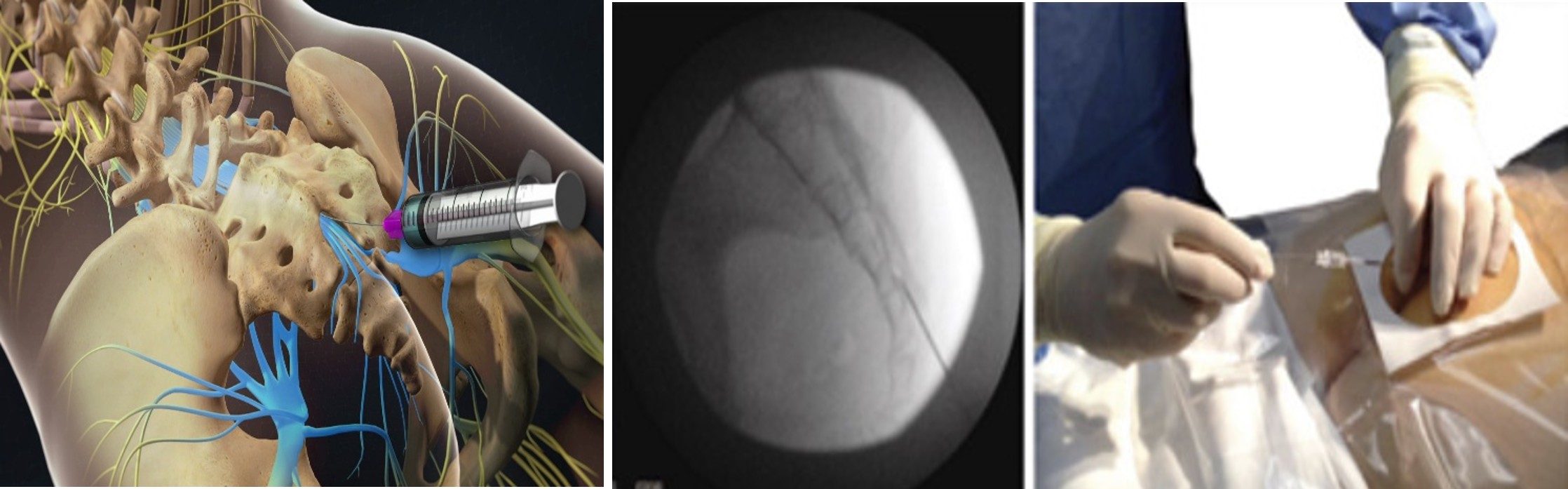
Caudal Epidural Steroid Injection (CESI) is a minimally invasive procedure in which corticosteroids combined with a local anesthetic are injected into the epidural space through the sacral hiatus under image guidance or anatomical landmark technique. The procedure reduces inflammation surrounding affected nerve roots, provides effective pain relief, improves functional recovery, and may delay or prevent surgical intervention. This case report describes the successful management of a young female patient with lumbar disc disease complicated by steroid-induced systemic adverse effects using caudal epidural steroid injection and highlights the essential role of multidisciplinary care and nursing management.
Case presentation
A 33-year-old female was brought to emergency department with complaints of severe low back pain, facial puffiness, and bilateral foot swelling for the past few days. The patient had initially received treatment at an outside hospital for similar complaints, where she was administered a steroid injection. Shortly after the injection, she developed facial swelling, bilateral lower limb edema, worsening back pain, and generalized discomfort. Due to deterioration of her clinical condition, she was referred to our hospital for further management.
Clinical Findings
On admission, the patient appeared uncomfortable because of severe pain.
Vital Signs
- HR – 43/min
- BP – 120/80 mmHg
- RR – 20/min
- Temperature – 97°F
- SpO2 – 94% on Room Air
- Supplemental oxygen initiated immediately
- Continuous cardiac monitoring commenced
Physical Examination
- CVS – S1S2 (+)
- P/A – Soft, non-tenderness
- CNS – NFND
- RS – B/L AE (+)
- Severe tenderness over the lumbar region
- Restricted lumbar movements
- Facial puffiness
- Bilateral pedal edema
- Radicular pain radiating to the lower limbs
- No bowel or bladder dysfunction
- No acute neurological deficit
Pain assessment using the Visual Analogue Scale (VAS) demonstrated a score of 8/10.
Investigations
| Test | Result |
|---|---|
| Hb | 11.3g/dl |
| Total Count | 17900/cumm |
| Platelet Count | 329000/cumm |
| Sodium | 140mmol/L |
| Potassium | 3.8mmol/L |
| Urea | 44.9 mg/dl |
| Creatinine | 1.0 mg/dl |
| Total Bilirubin | 0.5 mg/dl |
| Direct Bilirubin | 0.3 mg/dl |
| Indirect Bilirubin | 0.2 mg/dl |
| SGOT | 35 U/L |
| SGPT | 78 U/L |
| TSH | 2.023 mIU/L |
| T4 | 7.28 µg/dL |
| T3 | 0.705 ng/dl |
Echocardiogram (ECHO)
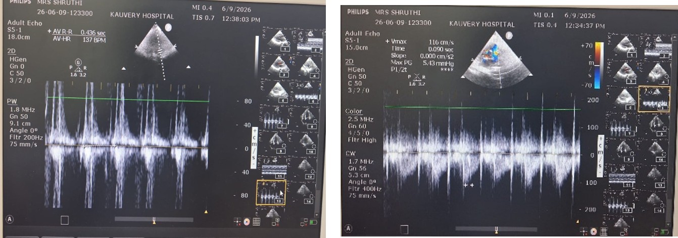
- Sinus tachycardia during study (Heart Rate: 137 bpm)
- Normal left ventricular function (LVEF 62%)
- Mild mitral regurgitation
- Trivial tricuspid regurgitation
Ultrasound Abdomen & Pelvis
- Liver: Grade I fatty liver
- Gallbladder: Contracted
- Right and Left Kidneys: Normal
- Bladder: Diffuse bladder wall thickening
- Small 8 mm umbilical hernia containing preperitoneal fat
MRI Lumbar Spine
- Mild lumbar spondylotic (degenerative) changes
- Transitional lumbosacral junction with sacralization of L5
- L4–L5:
-
- Disc desiccation (disc dehydration)
- Broad-based posterior central disc protrusion
- Small annular tear
- Indentation of the thecal sac
- Impingement of both traversing nerve roots at the lateral recess
- L3–L4:
-
- Mild posterior disc bulge causing mild indentation of the thecal sac and nerve root impingement
- Sacroiliac joints: Normal
MRI Cervical Spine
- C4–C5 and C5–C6:
-
- Mild posterior disc bulge
- Small posterolateral osteophyte (bone spur)
- Mild indentation of the thecal sac
- Minimal foraminal narrowing
- Mild posterior bulges also noted at the upper thoracic levels.
Diagnosis
The patient was diagnosed with:
- Lumbar Disc Disease with Lumbosacral Radiculopathy
- Steroid-Induced Fluid Retention
- Symptomatic Bradycardia
- Severe Chronic Low Back Pain
Nursing Management
Preoperative Nursing Management
- Recording baseline vital signs (blood pressure, heart rate, respiratory rate, temperature, and oxygen saturation).
- Assessing pain intensity using the Visual Analogue Scale (VAS).
- Performing a detailed neurological assessment, including motor strength, sensory function, and deep tendon reflexes of the lower extremities.
- Reviewing the patient’s medical history, current medications, and any previous adverse reactions, particularly to corticosteroids or local anaesthetics.
- Assessing for allergies to medications, antiseptic solutions, latex, or contrast agents (if applicable).
- Verifying fasting (NPO) status when indicated, in accordance with institutional protocol.
- Verification of the patient’s identity, procedure details, and informed written consent.
- Providing a clear explanation of the procedure, expected benefits, potential risks, and post-procedure care to reduce anxiety and improve cooperation.
- Offering emotional support and reassurance to alleviate procedural apprehension.
- Establishing and securing a patent intravenous (IV) access for medication administration and emergency management if required.
- Ensuring the bladder was emptied prior to the procedure to enhance patient comfort and minimize interruptions.
- Preparing emergency resuscitation equipment, medications, and monitoring devices to ensure immediate availability in the event of an adverse reaction.
- Confirming the availability of all sterile instruments, medications, and procedural supplies before transferring the patient to the procedure room.
Intra procedure Management
After obtaining informed written consent, the patient was transferred to the procedure room and standard intraoperative monitoring was established, including continuous electrocardiography (ECG), non-invasive blood pressure (NIBP), heart rate, and pulse oximetry. Intravenous access was secured, and oxygen supplementation was provided throughout the procedure. Strict aseptic precautions were maintained.
The patient was positioned in the prone position with a pillow placed under the abdomen to facilitate access to the sacral hiatus. Following skin preparation and sterile draping, the sacral hiatus was identified using anatomical landmarks. An epidural needle was carefully advanced through the sacral hiatus into the caudal epidural space. Correct needle placement was confirmed using standard procedural techniques.
A combination of corticosteroid and local anesthetic was then administered slowly into the epidural space after negative aspiration for blood or cerebrospinal fluid. The injection was delivered without resistance, and the patient remained hemodynamically stable throughout the procedure.
Continuous monitoring was maintained during and after the injection for any immediate adverse reactions or complications. The patient tolerated the procedure well, with no evidence of neurological deficits, allergic reactions, vascular injections, or other procedure-related complications. Following an appropriate observation period, the patient was transferred to the recovery area in stable conditions for further postoperative monitoring.
Post-Procedure Nursing Management
1. Vital Signs Monitoring
- The patient’s vital signs, including heart rate, blood pressure, respiratory rate, oxygen saturation, and temperature, were monitored regularly.
- The injection site was assessed for bleeding, hematoma formation, or signs of infection.
- The patient was observed closely for any immediate adverse reactions following the procedure.
2. Neurological Assessment
- Neurological status was assessed at regular intervals.
- Motor strength, sensory function, lower limb movement, and reflexes were evaluated.
- Bladder and bowel function were monitored to identify any signs of neurological compromise.
3. Pain Management
- Pain intensity was assessed using the Visual Analogue Scale (VAS).
- The patient’s response to the procedure was documented.
- Persistent or worsening pain was promptly communicated to the treating consultant, and appropriate pain management interventions were implemented as prescribed.
4. Positioning
- The patient was maintained in the supine position for 2–4 hours, as per institutional protocol.
- Gradual mobilization was initiated after the observation period, depending on the patient’s clinical condition.
5. Monitoring for Complications
- The patient was monitored for procedure-related complications, including headache, dizziness, dural puncture, infection, bleeding, allergic reactions, increased pain, or new neurological deficits.
- Any abnormal findings or warning signs were immediately reported to the treating physician for timely intervention.
6. Fluid Balance Monitoring
- Strict intake and output were recorded and monitored.
- Facial puffiness and bilateral pedal edema were assessed regularly to evaluate the resolution of steroid-induced fluid retention.
- The patient’s hydration status and overall fluid balance were monitored throughout the recovery period.
7. Patient Education
- The patient was educated regarding prescribed medications, activity restrictions, and the importance of adhering to physiotherapy.
- Instructions were provided regarding adequate fluid intake, follow-up appointments, and warning signs such as increasing pain, weakness, numbness, fever, urinary or bowel dysfunction, or persistent swelling that required immediate medical attention.
- The patient demonstrated understanding of the discharge instructions before leaving the hospital.
Outcome
The patient’s lumbar radicular pain was markedly reduced, with the Visual Analogue Scale (VAS) score improving from 8/10 at admission to 2/10 within 72 hours of the procedure. She experienced progressive relief of pain, resulting in improved mobility and the ability to perform daily activities with minimal discomfort. Facial puffiness and bilateral pedal edema, secondary to steroid-induced fluid retention, gradually resolved with conservative management and close monitoring. The patient’s heart rate improved, and bradycardia remained stable under conservative cardiology management without requiring further intervention. No neurological deficits, bowel or bladder dysfunction, or procedure-related complications such as dural puncture, infection, bleeding, or allergic reactions were observed. The patient tolerated the procedure well and remained hemodynamically stable throughout the post-procedure period. She actively participated in physiotherapy and demonstrated good compliance with the prescribed treatment plan. The patient was discharged in a stable clinical condition with significant pain relief, improved functional status, and comprehensive discharge education regarding medications, physiotherapy, and follow-up care. Overall, the patient expressed a high level of satisfaction with the treatment and reported a substantial improvement in her quality of life following the intervention.
Discharge
At the time of discharge, the patient was conscious, alert, and fully oriented to time, place, and person. She was hemodynamically stable, with vital signs within normal limits, and was maintaining adequate oxygen saturation on room air without the need for supplemental oxygen. Her lumbar radicular pain had significantly improved following the caudal epidural steroid injection, allowing her to ambulate independently with minimal discomfort. Neurological examination revealed intact motor and sensory functions, with no evidence of bowel or bladder dysfunction or any procedure-related complications. Facial puffiness and bilateral pedal edema had markedly reduced with conservative management. The patient was discharged in a stable clinical condition with instructions to continue the prescribed medications, participate in regular physiotherapy, adhere to spinal precautions, avoid heavy lifting and strenuous activities, and attend scheduled follow-up appointments with the Pain Management and Orthopedic teams for ongoing evaluation and rehabilitation.
Discussion – Medical Aspects
Lumbar disc disease is among the most common causes of lumbosacral radiculopathy. Persistent inflammation around compressed nerve roots results in severe pain and functional impairment. While conservative therapy remains the first-line treatment, patients with persistent symptoms may benefit from epidural steroid injections. Caudal Epidural Steroid Injection provides targeted delivery of corticosteroids into the epidural space, reducing inflammation and nerve root edema while minimizing systemic steroid exposure. Compared with repeated systemic corticosteroid administration, CESI offers localized therapeutic benefits with fewer systemic adverse effects.
In the present case, the patient developed steroid-induced fluid retention following systemic steroid administration, making further systemic corticosteroid therapy less desirable. After multidisciplinary evaluation involving emergency physicians, cardiologists, pain specialists, and nursing professionals, CESI was selected as an effective alternative. The procedure produced significant pain relief without worsening systemic complications. This case demonstrates that carefully selected interventional pain management can be safely performed even in patients who have experienced adverse reactions to previous systemic steroid therapy.
Discussion – Nursing Aspects
Nurses play a vital role throughout the peri-procedural management of patients undergoing Caudal Epidural Steroid Injection. Pre-procedure nursing responsibilities include comprehensive assessment, baseline neurological examination, pain assessment, allergy screening, patient education, informed consent verification, and psychological support to reduce anxiety. During the procedure, nurses assist in maintaining aseptic technique, monitoring vital signs, preparing emergency equipment, and ensuring patient safety. Post-procedure care focuses on early identification of complications through continuous monitoring of vital signs, neurological status, puncture site assessment, pain evaluation, and fluid balance monitoring. Accurate documentation and timely communication with the medical team facilitate prompt intervention when necessary. Patient education regarding medication adherence, physiotherapy, activity modification, warning signs, and follow-up care is essential for preventing recurrence and promoting long-term recovery. The coordinated efforts of nursing professionals significantly contributed to the patient’s safe recovery, improved comfort, and high level of satisfaction with care.
Conclusion
Caudal Epidural Steroid Injection is a safe, effective, and minimally invasive intervention for patients with lumbar radicular pain who do not respond adequately to conservative management. The procedure provides rapid pain relief, improves functional recovery, and may reduce the need for surgical intervention. This case demonstrates that even in patients who develop systemic adverse effects following corticosteroid administration, carefully planned interventional pain management can be successfully performed with favourable clinical outcomes. Comprehensive nursing care, multidisciplinary collaboration, vigilant monitoring, and patient education are essential components of successful treatment and recovery.
Learning Points
- Caudal Epidural Steroid Injection is an effective alternative to surgery for selected patients with lumbar radiculopathy.
- Early multidisciplinary decision-making improves patient safety and outcomes.
- Comprehensive nursing assessment and monitoring are essential before and after the procedure.
- Patient education and physiotherapy play a key role in preventing recurrence and improving long-term functional recovery.
- Careful monitoring for systemic steroid-related adverse effects should guide individualized treatment planning.