
Acute type A aortic dissection in a patient with marfanoid habitus, aortic root aneurysm, and mitral valve prolapse: A comprehensive case report with anatomical, pathophysiological, surgical, and nursing perspectives
Gowdham Pannirselvam1*, Shilpa S2
1Senior Nurse Educator, Kauvery Hospital, Marathahalli, Bangalore
2In charge CT-ICU, Kauvery Hospital, Marathahalli, Bangalore
*Correspondence
Abstract
Acute Type A aortic dissection is among the most catastrophic cardiovascular emergencies, associated with significant morbidity and mortality despite advances in diagnostic modalities and surgical management. The condition frequently occurs in individuals with underlying connective tissue disorders, particularly Marfan syndrome, where structural weakness of the aortic wall predisposes patients to progressive aortic dilatation, aneurysm formation, and eventual dissection. This case describes a 40-year-old female with marfanoid habitus, known mitral valve prolapses, and aortic root aneurysm who presented with acute onset breathlessness, vomiting, giddiness, and right shoulder pain. Clinical and radiological evaluation revealed an acute Type A aortic dissection involving the aortic root aneurysm. The patient underwent emergency Bentall procedure with mitral valve repair and coronary artery bypass grafting. Despite aggressive postoperative management involving mechanical ventilation, intra-aortic balloon pump support, multiple inotropes, blood product transfusions, and continuous renal replacement therapy, the patient developed progressive cardiogenic shock and multiorgan dysfunction syndrome, culminating in death. This explores the anatomical and physiological basis of the disease, the pathophysiological mechanisms underlying the patient’s deterioration, surgical decision-making, intensive care management, and nursing implications. The case highlights the importance of early diagnosis, routine surveillance of aortic pathology in high-risk patients, and multidisciplinary management in improving patient outcomes.
Keywords: Acute Type A aortic dissection; Marfan syndrome; Aortic root aneurysm; Bentall procedure; Mitral valve prolapse; Cardiogenic shock; Multiorgan dysfunction syndrome.
Introduction
Acute aortic dissection represents one of the most life-threatening cardiovascular emergencies encountered in clinical practice. It occurs when a tear develops in the intimal layer of the aortic wall, permitting blood to enter the medial layer and create a false lumen that separates the layers of the vessel wall. Depending on the location of involvement, dissections are classified using the Stanford classification system into Type A and Type B dissections. Stanford Type A dissections involve the ascending aorta and constitute surgical emergencies because mortality increases dramatically with every hour of delay in treatment. The incidence of acute aortic dissection is estimated to range between 3 and 6 cases per 100,000 population annually. Although hypertension remains the most common risk factor in older individuals, connective tissue disorders such as Marfan syndrome significantly increase the risk among younger patients. Marfan syndrome is an autosomal dominant connective tissue disorder caused primarily by mutations in the fibrillin-1 (FBN1) gene. Defective fibrillin results in weakening of elastic connective tissue throughout the body, particularly affecting the cardiovascular, skeletal, and ocular systems. Cardiovascular manifestations are responsible for most deaths associated with Marfan syndrome and include aortic root dilatation, aortic aneurysm formation, aortic dissection, and mitral valve prolapse. Without appropriate surveillance and intervention, progressive enlargement of the aortic root may culminate in catastrophic dissection or rupture. The present case describes a young female with marfanoid habitus and known aortic root aneurysm who developed an acute Type A aortic dissection requiring emergency surgery. Despite successful operative correction of the anatomical defect, she developed refractory cardiogenic shock and multiorgan dysfunction syndrome, ultimately resulting in death. This case provides an opportunity to review the complex interplay between connective tissue disorders, aortic pathology, surgical intervention, intensive care management, and nursing care.
Case presentation
A 40-year-old female presented to the emergency department with complaints of sudden onset breathlessness, repeated episodes of vomiting, giddiness, and right shoulder pain that had begun a few hours prior to admission. According to the available clinical documentation, she was a known case of mitral valve prolapse and had been under follow-up for an aortic root aneurysm that had been identified several years earlier. The patient also had a history of total thyroidectomy performed approximately four years previously for carcinoma thyroid and was receiving suppressive therapy with thyroxine. Clinical notes documented the presence of marfanoid habitus, suggesting an underlying connective tissue disorder. Marfanoid habitus refers to characteristic skeletal features including tall stature, long limbs, elongated fingers, increased arm-span-to-height ratio, and other manifestations commonly associated with Marfan syndrome. Such findings are important because they often provide the first clinical clue to the presence of potentially life-threatening cardiovascular abnormalities.
At presentation, the patient was categorized as New York Heart Association (NYHA) Class III, indicating significant limitation of physical activity due to cardiac symptoms. Initial evaluation raised concern for progression of the previously known aortic root aneurysm. Imaging investigations, including CT angiography, demonstrated an acute Type A aortic dissection involving the ascending aorta and aortic root aneurysm. The diagnosis represented a cardiovascular emergency because Type A dissections are associated with high rates of mortality due to rupture, cardiac tamponade, coronary artery compromise, severe aortic regurgitation, and cardiogenic shock. After detailed counselling regarding risks and benefits, the patient underwent emergency surgical intervention. The operative procedure included a Bentall procedure utilizing a 23-mm St. Jude mechanical valve conduit and a 26-mm Dacron graft for replacement of the diseased aortic root and ascending aorta. Simultaneously, mitral valve repair was performed because of the pre-existing mitral valve prolapse. Coronary artery bypass grafting using a reversed saphenous vein graft to the left anterior descending artery was also undertaken. Following surgery, the patient was transferred to the intensive care unit for advanced hemodynamic monitoring and organ support. Initially, there appeared to be transient improvement in her condition. However, over the ensuing days she developed progressive hemodynamic instability requiring escalating doses of inotropic agents, intra-aortic balloon pump support, blood transfusions, ventilatory support, and continuous renal replacement therapy. Despite maximal intensive care measures, her condition continued to deteriorate, and she ultimately succumbed to cardiogenic shock and multiorgan dysfunction syndrome.
Anatomy and Physiology of the Aortic Root
The aortic root represents the initial segment of the aorta and forms the anatomical junction between the left ventricle and the systemic circulation. It is a highly specialized structure designed not only to transport blood but also to facilitate efficient valvular function and coronary artery perfusion. Anatomically, the aortic root extends from the aortic annulus to the Sino tubular junction and includes the aortic valve leaflets, the sinuses of Valsalva, and the origins of the coronary arteries. The normal diameter of the aortic root generally ranges between 2.0 and 3.7 cm depending upon age, sex, and body surface area.
The aortic wall itself is composed of three distinct layers: the intima, media, and adventitia. The intima forms the innermost endothelial lining and provides a smooth, non-thrombogenic surface for blood flow. The media constitutes the thickest layer and contains concentric layers of elastic fibres, collagen fibres, and smooth muscle cells. This layer is responsible for maintaining the structural integrity and elasticity of the vessel. The outermost adventitia contains connective tissue, fibroblasts, collagen fibres, lymphatics, and the vasa vasorum that supply nutrients to the aortic wall.
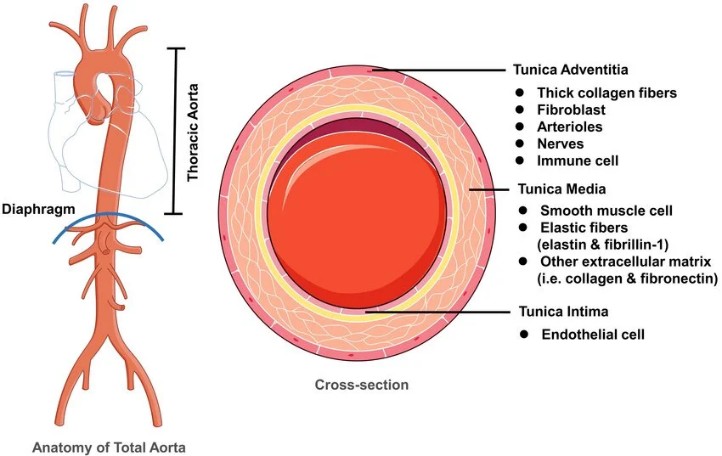
Image Source: AI Image
The physiological function of the aortic root extends beyond simple blood conduction. During ventricular systole, blood is ejected from the left ventricle through the aortic valve into the aorta. The elastic nature of the aortic wall allows it to expand and accommodate the stroke volume. During diastole, elastic recoil of the aortic wall helps maintain continuous blood flow to peripheral tissues. This phenomenon, known as the Wind Kessel effect, reduces fluctuations in arterial pressure and improves perfusion of vital organs. Another critical function of the aortic root is facilitation of coronary circulation. Blood enters the coronary arteries during diastole through the coronary ostia located within the right and left sinuses of Valsalva. Any pathological process affecting the aortic root, such as aneurysm formation or dissection, may compromise coronary perfusion and result in myocardial ischemia or infarction. In the present case, the patient had a known aortic root aneurysm. Progressive enlargement of the aortic root significantly weakens the vessel wall and increases the likelihood of intimal tearing, eventually leading to aortic dissection. This pathological alteration represents one of the most feared cardiovascular complications in patients with connective tissue disorders such as Marfan syndrome.
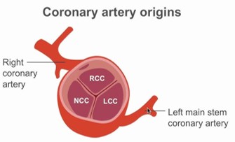
Image Source: AI Image
Anatomy and Physiology of the Ascending Aorta
The ascending aorta begins at the Sino tubular junction and extends superiorly until it reaches the aortic arch. It measures approximately 5–8 cm in length and serves as the primary conduit for transporting oxygenated blood from the heart to the systemic circulation. Structurally, the ascending aorta contains a high concentration of elastic fibers within its medial layer. These elastic fibers permit repeated expansion and recoil during each cardiac cycle. This elasticity is essential because the ascending aorta is exposed to the highest pressures generated by the left ventricle. Under normal circumstances, the ascending aorta can withstand substantial hemodynamic stress while maintaining structural integrity. The ascending aorta plays a central role in systemic hemodynamic. During ventricular contraction, the vessel expands and stores mechanical energy. During ventricular relaxation, the stored energy is released, promoting forward blood flow and maintaining arterial pressure. This mechanism significantly reduces cardiac workload and enhances circulatory efficiency.
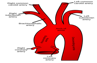
Image Source: AI Image
In patients with connective tissue disorders, degeneration of elastic fibers within the media weakens the vessel wall. Over time, the ascending aorta becomes progressively dilated and susceptible to aneurysm formation. As vessel diameter increases, wall tension rises according to Laplace’s law, thereby increasing the risk of dissection and rupture. In this patient, chronic dilation of the ascending aorta eventually culminated in acute Type A dissection. Once the intimal tear occurred, blood entered the medial layer and propagated longitudinally along the vessel wall, creating a false lumen. This process disrupted normal vascular architecture and compromised blood flow to vital organs.
Anatomy and Physiology of the Mitral Valve
The mitral valve is located between the left atrium and left ventricle and functions as the primary regulator of blood flow between these chambers. It is composed of two leaflets, the anterior leaflet and posterior leaflet, supported by chordae tendineae and papillary muscles. Together, these structures form a highly coordinated valvular apparatus that ensures unidirectional blood flow. During ventricular diastole, the mitral valve opens, allowing oxygenated blood from the left atrium to flow into the left ventricle. During ventricular systole, contraction of the papillary muscles maintains tension on the chordae tendineae, preventing prolapse of the valve leaflets into the left atrium. Effective closure of the valve prevents regurgitation and ensures efficient forward cardiac output. The mitral valve is particularly vulnerable to connective tissue abnormalities. In Marfan syndrome, myxomatous degeneration weakens the valve leaflets and supporting structures. As a result, one or both leaflets may bulge backward into the left atrium during systole, producing mitral valve prolapse (MVP).
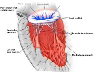
Image Source: AI Image
Mitral valves prolapse is among the most common cardiovascular manifestations of Marfan syndrome. Although many patients remain asymptomatic, progressive prolapse may lead to significant mitral regurgitation, atrial enlargement, arrhythmias, heart failure, and increased mortality. The patient had a known history of mitral valve prolapse. The coexistence of MVP and aortic root aneurysm strongly supports the likelihood of an underlying connective tissue disorder affecting multiple cardiovascular structures simultaneously.
Anatomy and Physiology of the Coronary Arteries
The coronary arteries arise from the aortic root immediately above the aortic valve within the sinuses of Valsalva. The right coronary artery originates from the right coronary sinus, while the left main coronary artery arises from the left coronary sinus and subsequently divides into the left anterior descending artery and circumflex artery. The coronary circulation supplies oxygen and nutrients to the myocardium. Unlike most organs, myocardial perfusion occurs predominantly during diastole because systolic contraction compresses intramyocardial vessels. Consequently, adequate diastolic pressure within the aortic root is essential for maintaining coronary blood flow.
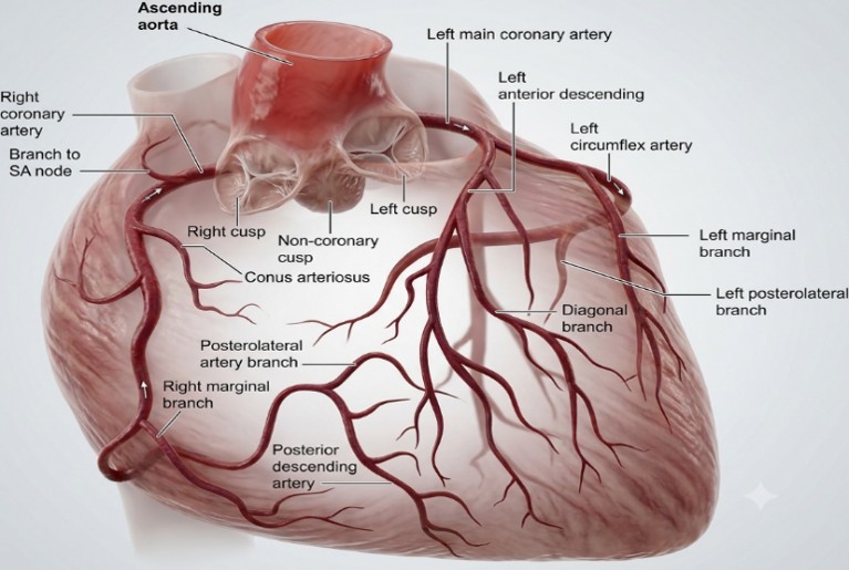
Image Source: AI Image
Acute aortic dissection involving the ascending aorta may extend into the coronary ostia. When this occurs, coronary blood flow becomes compromised, resulting in myocardial ischemia, ventricular dysfunction, arrhythmias, and cardiogenic shock. Coronary Malperfusion represents one of the most important predictors of mortality in acute Type A dissection. In this case, coronary artery involvement likely contributed to the decision to perform coronary artery bypass grafting during surgical repair. Bypass grafting ensures adequate myocardial perfusion when coronary artery anatomy is distorted or compromised by the dissection process.
Anatomical Basis of Marfanoid Habitus
The term “marfanoid habitus” describes a collection of skeletal features suggestive of an underlying connective tissue disorder. These characteristics arise because fibrillin, a major component of connective tissue microfibrils, is widely distributed throughout the body. Defective fibrillin production affects the musculoskeletal, cardiovascular, and ocular systems. Common skeletal manifestations include increased height, disproportionately long extremities, arachnodactyly, pectus deformities, scoliosis, joint hypermobility, and a high-arched palate. Although these features may initially appear benign, their presence often serves as an important clinical marker of potentially life-threatening cardiovascular disease. In the present patient, marfanoid habitus combined with mitral valve prolapse and aortic root aneurysm strongly suggested an underlying connective tissue disorder like Marfan syndrome. Recognition of these features is crucial because regular surveillance of aortic dimensions and timely prophylactic surgery can significantly reduce the risk of fatal aortic dissection.
Relationship Between Anatomy and Clinical Presentation
The patient’s symptoms can be directly explained by the anatomical structures affected by the disease process. The sudden onset of breathlessness likely reflected acute cardiac dysfunction resulting from compromised ventricular performance and altered hemodynamic. Right shoulder pain may have resulted from extension of the dissection, irritation of adjacent mediastinal structures, or referred pain pathways. Vomiting and giddiness likely reflected reduced systemic perfusion and activation of autonomic responses secondary to acute cardiovascular collapse. The coexistence of aortic root aneurysm, mitral valve prolapses, and marfanoid habitus indicates a systemic connective tissue abnormality affecting multiple cardiovascular structures simultaneously. Progressive weakening of the aortic wall eventually culminated in dissection, creating a catastrophic disruption of normal cardiovascular anatomy and physiology. Understanding these anatomical relationships is essential for appreciating the rapid clinical deterioration observed in this patient.
Pathophysiology of Marfan Syndrome
Marfan syndrome is an autosomal dominant connective tissue disorder caused primarily by mutations in the fibrillin-1 (FBN1) gene located on chromosome 15. Fibrillin is an essential glycoprotein responsible for the formation and maintenance of elastic fibers within connective tissues throughout the body. Elastic fibers provide strength, elasticity, and structural support to various organs, particularly the cardiovascular system, skeletal system, ocular structures, and blood vessels. Under normal physiological conditions, fibrillin microfibrils serve as scaffolding for elastin deposition and regulate transforming growth factor-beta (TGF-β) activity. In patients with Marfan syndrome, defective fibrillin results in loss of structural integrity and abnormal TGF-β signalling. Increased TGF-β activity promotes degradation of extracellular matrix components, apoptosis of smooth muscle cells, and progressive weakening of connective tissues. The cardiovascular system is particularly susceptible because the aortic wall relies heavily on elastic fibers to withstand continuous hemodynamic stress. Progressive destruction of elastic fibers within the tunica media leads to cystic medial degeneration, characterized by fragmentation of elastic fibers, accumulation of mucoid material, and loss of smooth muscle cells. These pathological changes gradually weaken the aortic wall and predispose patients to aneurysm formation and dissection.
The patient’s medical conditions such as marfanoid habitus, mitral valve prolapse, and aortic root aneurysm strongly suggest the presence of an underlying connective tissue disorder resembling Marfan syndrome. The coexistence of these findings demonstrates how a single genetic defect can affect multiple organ systems simultaneously. Although skeletal manifestations often attract initial attention, cardiovascular complications remain the leading cause of morbidity and mortality in these patients. Without early identification and surveillance, progressive weakening of the aortic wall may continue silently for years before presenting catastrophically as acute aortic dissection. Therefore, recognition of marfanoid features should always prompt comprehensive cardiovascular evaluation, including echocardiography and advanced vascular imaging.
Pathophysiology of Aortic Root Aneurysm
An aortic aneurysm is defined as a localized dilatation of the vessel exceeding 50% of its normal diameter. In patients with connective tissue disorders, aneurysm formation is primarily driven by structural weakness of the medial layer of the aortic wall. The normal aortic wall contains highly organized elastic lamellae that distribute mechanical stress evenly throughout the vessel. In Marfan syndrome, fragmentation of these elastic fibers progressively reduces the tensile strength of the aorta. As a result, the vessel becomes unable to withstand normal systolic pressures and gradually enlarges over time.
The aortic root is particularly vulnerable because it experiences substantial mechanical stress during every cardiac cycle. Repeated exposure to pulsatile pressure accelerates degeneration of the already weakened vessel wall. Enlargement of the aortic root creates a vicious cycle in which increasing diameter leads to increasing wall tension according to Laplace’s law.
T = (P × r) / (2h)
Where:
- T = Wall tension
- P = Intraluminal pressure
- r = Radius of the vessel
- h = Wall thickness
As the radius increases, wall tension rises proportionally, further accelerating aneurysm expansion and increasing the likelihood of rupture or dissection.
In the present case, the patient had an aortic root aneurysm under follow-up for approximately ten years. The prolonged duration suggests gradual progressive enlargement of the aortic root until the vessel wall eventually reached a critical point of structural failure. The aneurysm served as the anatomical substrate upon which acute dissection subsequently developed. Another important consequence of aortic root aneurysm is distortion of the aortic valve apparatus. Enlargement of the aortic annulus may prevent proper coaptation of valve leaflets, leading to aortic regurgitation. Severe aortic regurgitation increases left ventricular workload and contributes to ventricular dysfunction, pulmonary congestion, and heart failure.
Pathophysiology of Mitral Valve Prolapse
Mitral valve prolapse (MVP) is one of the most common cardiovascular manifestations of Marfan syndrome. It occurs when one or both mitral valve leaflets bulge abnormally into the left atrium during ventricular systole. The condition results primarily from myxomatous degeneration of the valve leaflets and supporting structures. In normal physiology, the mitral valve closes tightly during ventricular contraction, preventing backward flow of blood into the left atrium. Effective valve closure depends upon the coordinated function of the leaflets, chordae tendineae, papillary muscles, and mitral annulus.
In connective tissue disorders, weakening of collagen and elastin fibers leads to thickening, elongation, and redundancy of the valve leaflets. The chordae tendineae may also become elongated and fragile. Consequently, the valve leaflets prolapse into the left atrium during systole and fail to achieve complete closure.
Initially, MVP may be asymptomatic. However, progressive prolapse often leads to mitral regurgitation, causing chronic volume overload of the left atrium and left ventricle. Over time, this results in chamber enlargement, arrhythmias, pulmonary hypertension, reduced exercise tolerance, and heart failure. In this patient, mitral valve prolapses had been recognized before presentation and was significant enough to warrant surgical repair during the Bentall procedure. The coexistence of MVP and aortic root aneurysm reflects the systemic nature of connective tissue involvement and contributed to the overall complexity of surgical management.
Pathophysiology of Acute Type A Aortic Dissection
Acute aortic dissection occurs when an intimal tear allows blood to penetrate the medial layer of the aortic wall. The high-pressure blood column then separates the layers of the vessel, creating a false lumen that may extend both proximally and distally. In this patient, chronic degeneration of the aortic wall likely resulted in an intimal tear within the ascending aorta. Once blood entered the media, propagation of the dissection occurred rapidly due to the high pressures generated by left ventricular contraction.
The pathological sequence begins with intimal disruption. Blood then dissects through weakened medial tissue planes, producing two channels: the original true lumen and a newly formed false lumen. As the false lumen expands, it may compress the true lumen and compromise blood flow to vital organs.
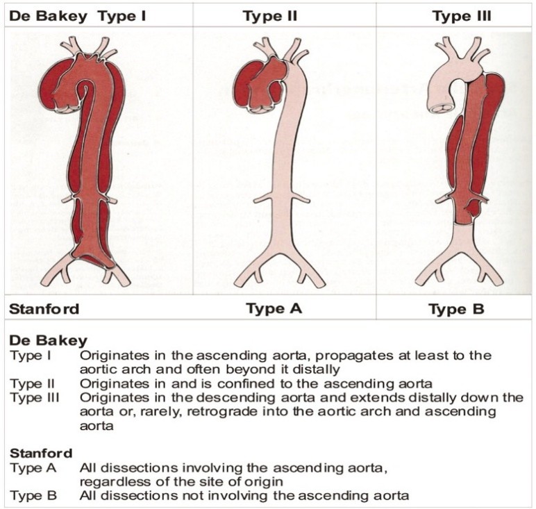
Image Source: AI Image
Stanford Type A dissections are particularly dangerous because they involve the ascending aorta. Several life-threatening complications may occur simultaneously:
Coronary Artery Involvement
The coronary arteries originate from the aortic root. Extension of the dissection into the coronary ostia may obstruct coronary blood flow and cause acute myocardial ischemia or infarction. Myocardial dysfunction resulting from coronary malperfusion is a major contributor to cardiogenic shock.
Acute Aortic Regurgitation
When the dissection extends into the aortic root, separation of supporting structures may prevent normal valve closure. Sudden severe aortic regurgitation develops, causing rapid volume overload of the left ventricle. The ventricle is unable to adapt acutely, resulting in pulmonary edema and severe hemodynamic compromise.
Cardiac Tamponade
Dissection may rupture through the adventitia into the pericardial space, causing accumulation of blood around the heart. Cardiac tamponade impairs ventricular filling and may lead to sudden circulatory collapse.
Organ Malperfusion
The false lumen may obstruct major arterial branches supplying the brain, kidneys, intestines, liver, or extremities. Organ ischemia dramatically worsens prognosis and contributes to multiorgan failure. The patient’s symptoms of sudden breathlessness, vomiting, giddiness, and right shoulder pain were entirely consistent with acute aortic dissection and systemic hypoperfusion. By the time surgical intervention was undertaken, significant physiological injury had likely already occurred.
Development of Cardiogenic Shock
Cardiogenic shock is a state of inadequate tissue perfusion resulting from severe cardiac dysfunction. In this patient, multiple mechanisms acted simultaneously to impair cardiac output.
First, coronary malperfusion associated with the dissection may have reduced myocardial blood supply. Ischemic myocardium loses its contractile ability, reducing stroke volume and cardiac output.
Second, acute aortic regurgitation increases left ventricular end-diastolic volume and pressure. The ventricle becomes unable to maintain effective forward flow, resulting in pulmonary congestion and systemic hypoperfusion.
Third, major cardiac surgery itself imposes substantial physiological stress. Cardiopulmonary bypass, myocardial ischemia-reperfusion injury, systemic inflammatory activation, and postoperative ventricular dysfunction may all contribute to shock.
The patient status indicate that the patient required multiple inotropic agents and intra-aortic balloon pump (IABP) support. These interventions suggest severe myocardial dysfunction and persistent circulatory instability despite aggressive treatment. As cardiac output declined, oxygen delivery to tissues became inadequate. Cellular metabolism shifted from aerobic to anaerobic pathways, leading to lactic acidosis, endothelial injury, and progressive organ dysfunction.
Pathophysiology of Multiorgan Dysfunction Syndrome (MODS)
Multiorgan dysfunction syndrome represents the final common pathway of severe shock and systemic inflammation. It occurs when dysfunction develops in two or more organ systems such that normal homeostasis cannot be maintained without therapeutic intervention.
In this patient, cardiogenic shock initiated a cascade of events that ultimately culminated in MODS. Persistent tissue hypoperfusion triggered widespread cellular injury, inflammatory cytokine release, endothelial dysfunction, and microvascular thrombosis.
Renal Dysfunction
The kidneys are highly sensitive to reductions in blood flow. Prolonged hypotension resulted in acute kidney injury requiring continuous renal replacement therapy (CRRT). Renal dysfunction further aggravated fluid overload, electrolyte disturbances, and metabolic derangements.
Hepatic Dysfunction
The patient had ischemic hepatitis. Reduced hepatic perfusion causes hepatocellular necrosis and marked elevation of liver enzymes. Hepatic failure also impairs coagulation factor synthesis, contributing to bleeding complications.
Haematological Dysfunction
Major surgery, massive transfusions, inflammation, and hepatic dysfunction disrupt normal coagulation mechanisms. The patient required repeated blood product support, indicating significant coagulopathy and bleeding risk.
Respiratory Dysfunction
Cardiogenic shock frequently results in pulmonary edema and acute respiratory failure. Mechanical ventilation was necessary to maintain oxygenation and reduce myocardial oxygen demand.
Ultimately, despite maximal supportive care, progressive organ failure became irreversible. The patient’s terminal deterioration was characterized by worsening shock, refractory hypotension, severe metabolic dysfunction, pulseless electrical activity, and unsuccessful cardiopulmonary resuscitation.
Transition to Surgical Management
The pathological sequence observed in this case illustrates the devastating progression from connective tissue disorder to aneurysm formation, acute dissection, cardiogenic shock, and multiorgan failure. Understanding these mechanisms is essential for appreciating the rationale behind emergency surgical intervention. The next section will examine the Bentall procedure, mitral valve repair, coronary artery bypass grafting, and the surgical principles that guided management of this complex case.
Surgical Decision-Making in Acute Type A Aortic Dissection
Acute Type A aortic dissection is considered one of the most challenging emergencies in cardiovascular surgery. Once the diagnosis is established, surgical intervention must be undertaken immediately because mortality increases by approximately 1–2% per hour during the first 24 hours after symptom onset. The primary objectives of surgery are to prevent aortic rupture, eliminate the intimal tear, restore normal blood flow through the true lumen, preserve coronary perfusion, and prevent life-threatening complications such as cardiac tamponade and severe aortic regurgitation.
In this case, the patient had multiple high-risk cardiovascular abnormalities including aortic root aneurysm, acute Type A dissection, mitral valve prolapse, and probable coronary artery involvement. These abnormalities required a complex surgical strategy rather than a simple replacement of the ascending aorta. The surgical team therefore opted for a Bentall procedure combined with mitral valve repair and coronary artery bypass grafting (CABG). The decision to perform such an extensive procedure reflects the severity of the anatomical damage identified during surgical evaluation. Merely repairing the dissected segment would not have addressed the underlying aortic root pathology or the mitral valve abnormality. Therefore, a comprehensive surgical approach was required to maximize the likelihood of survival.
Bentall Procedure: Historical Background
The Bentall procedure was first described by Hugh Bentall and Antony De Bono in 1968 as a method for treating combined disease of the ascending aorta and aortic valve. Since its introduction, the procedure has become the gold standard treatment for patients with aortic root aneurysms, aortic root dissections, connective tissue disorders, and complex aortic valve pathology.
The procedure involves replacement of:
- The diseased aortic root
- The ascending aorta
- The aortic valve
- Reimplantation of the coronary arteries
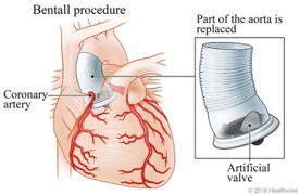
Image Source: AI Source
By replacing all diseased structures simultaneously, the Bentall procedure provides definitive treatment for extensive pathology involving the proximal aorta.
In patients with Marfan syndrome or connective tissue disorders, the Bentall procedure is particularly beneficial because the native aortic tissue is inherently weak and prone to recurrent aneurysm formation.
Anatomical Basis for the Bentall Procedure
To understand why the Bentall procedure was necessary in this patient, it is important to review the anatomical relationships involved.
The aortic root contains:
- Aortic annulus
- Aortic valve
- Sinuses of Valsalva
- Coronary artery origins
- Proximal ascending aorta
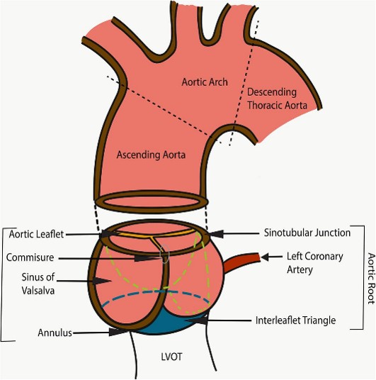
Image Source: AI Generated
In this patient, the dissection occurred within a previously enlarged aortic root aneurysm. Therefore, simply replacing the ascending aorta would have left behind diseased tissue capable of future rupture or recurrent dissection.
The Bentall procedure addressed all these issues by completely replacing the diseased root and ascending aorta while simultaneously restoring normal coronary circulation and valvular function.
Why Was a Mechanical Valve Used?
For, this patient we used a St. Jude mechanical valve.
Mechanical valves are often selected in younger patients because they offer exceptional durability and can function effectively for decades.
Advantages include:
- Excellent long-term durability
- Lower risk of structural valve degeneration
- Reduced likelihood of future reoperation
However, mechanical valves require lifelong anticoagulation therapy because of their thrombogenic potential.
In contrast, bioprosthetic valves avoid the need for long-term anticoagulation but undergo structural deterioration over time, particularly in younger individuals. Given the patient’s age of 40 years, implantation of a mechanical valve was an appropriate surgical decision aimed at maximizing long-term durability.
Mitral Valve Repair
Mitral valve prolapse was another major component of the patient’s cardiovascular pathology.
During surgery, the mitral valve was repaired rather than replaced.
Mitral valve repair is generally preferred over replacement because it:
- Preserves native valve anatomy
- Maintains ventricular geometry
- Reduces risk of prosthetic valve complications
- Improves long-term survival
- Reduces thromboembolic events
In this patient, repair was likely performed to correct leaflet prolapse and restore normal valve competence while preserving ventricular function.
Coronary Artery Bypass Grafting (CABG)
For This patient a reversed saphenous vein graft was used to bypass the left anterior descending artery.
Alternative Surgical Approaches
Several alternative procedures exist for management of aortic root pathology.
Valve-Sparing Root Replacement
The David procedure preserves the patient’s native aortic valve while replacing the aortic root.
Advantages include:
- Avoidance of prosthetic valve
- No lifelong anticoagulation
- Better hemodynamic performance
However, this approach is feasible only when the native valve leaflets remain structurally normal.
Because this patient had associated mitral valve pathology and acute dissection, a conventional Bentall procedure provided a safer and more definitive solution.
Isolated Ascending Aortic Replacement
In some cases, surgeons replace only the ascending aorta.
This approach is unsuitable when:
- Aortic root aneurysm is present
- Coronary ostia are involved
- Aortic valve pathology exists
Since all three conditions were likely present in this patient, isolated replacement would not have adequately treated the disease.
Immediate Postoperative Goals
Following completion of surgery, several critical objectives guide intensive care management.
These include:
- Maintaining adequate cardiac output
- Preventing myocardial ischemia
- Optimizing oxygen delivery
- Monitoring for bleeding
- Preserving organ perfusion
- Detecting neurological complications
- Supporting renal function
Initially, the patient demonstrated transient improvement. However, despite successful anatomical correction of the dissection, severe physiological injury continued to progress.
The need for:
- Triple inotropic support
- Intra-aortic balloon pump (IABP)
- Mechanical ventilation
- Blood transfusions
- Continuous renal replacement therapy
indicated severe postoperative cardiac dysfunction and evolving multiorgan failure.
Why Surgery Could Not Ultimately Save the Patient?
Although surgery successfully repaired the anatomical defects, it could not reverse the extensive physiological damage that had already occurred.
Several factors likely contributed:
The patient presented with an acute Type A dissection involving a large aortic root aneurysm. Significant myocardial ischemia may have developed before surgery because of compromised coronary circulation. Prolonged systemic hypoperfusion likely initiated cellular injury in multiple organs even before arrival in the operating room.
Furthermore, emergency cardiac surgery itself imposes substantial physiological stress. Cardiopulmonary bypass, ischemia-reperfusion injury, systemic inflammation, coagulopathy, and postoperative ventricular dysfunction all contribute to organ injury.
Consequently, while the anatomical problem was corrected successfully, irreversible physiological damage continued to progress, eventually leading to refractory cardiogenic shock and multiorgan dysfunction syndrome.
Postoperative Intensive Care Unit Course
Following completion of the Bentall procedure, mitral valve repair, and coronary artery bypass grafting, the patient was transferred to the cardiothoracic intensive care unit for advanced postoperative monitoring and organ support. The immediate postoperative period following repair of an acute Type A aortic dissection is often characterized by significant hemodynamic instability due to the combined effects of pre-existing cardiovascular compromise, prolonged cardiopulmonary bypass, myocardial ischemia-reperfusion injury, systemic inflammatory response, and major surgical trauma.
Initially, the primary goals of intensive care management were stabilization of hemodynamics, maintenance of adequate cardiac output, optimization of oxygen delivery, prevention of bleeding complications, and preservation of organ perfusion. Continuous invasive monitoring was established, including arterial pressure monitoring, central venous pressure monitoring, urine output measurement, serial arterial blood gas analysis, and frequent laboratory investigations.
Despite successful surgical correction of the anatomical abnormalities, the patient remained critically ill. Due to the condition, she required mechanical ventilation, multiple inotropic agents, and intra-aortic balloon pump (IABP) support. The requirement for escalating circulatory support suggested severe postoperative myocardial dysfunction and an inability of the heart to maintain adequate systemic perfusion independently.
The development of persistent hypotension despite maximal pharmacological support indicated progression toward cardiogenic shock. As cardiac output declined, oxygen delivery to vital organs became increasingly compromised. The resulting imbalance between oxygen supply and metabolic demand initiated a cascade of cellular injury that ultimately culminated in multiorgan dysfunction syndrome.
Development of Cardiogenic Shock
Cardiogenic shock represents a state of critical end-organ hypoperfusion caused by severe cardiac dysfunction. It is one of the most serious complications following acute Type A aortic dissection and remains a major cause of postoperative mortality.
Several mechanisms likely contributed to the development of cardiogenic shock in this patient. First, coronary artery involvement associated with the dissection may have caused significant myocardial ischemia before surgical intervention. Even when coronary blood flow is restored surgically, prolonged ischemia can result in irreversible myocardial injury and reduced ventricular contractility.
Second, acute aortic regurgitation caused by disruption of the aortic root may have imposed substantial volume overload on the left ventricle. Sudden increases in ventricular preload can rapidly impair myocardial performance and precipitate pulmonary edema.
Third, prolonged cardiopulmonary bypass and aortic cross-clamping are known to cause myocardial stunning. Myocardial stunning refers to transient but severe ventricular dysfunction occurring after restoration of blood flow following ischemia. Although potentially reversible, severe myocardial stunning can persist for several days and contribute significantly to postoperative shock.
Clinical evidence supporting severe cardiogenic shock in this patient includes the need for triple inotropic support and intra-aortic balloon pump assistance. These interventions are typically reserved for patients with profound circulatory failure who are unable to maintain adequate tissue perfusion despite conventional treatment.
Role of the Intra-Aortic Balloon Pump
The intra-aortic balloon pump is a mechanical circulatory support device commonly used in severe cardiogenic shock. The device functions through counter pulsation, inflating during diastole and deflating immediately before systole.
Inflation during diastole increases coronary artery perfusion by augmenting diastolic pressure within the aorta. Deflation before systole reduces left ventricular afterload, thereby decreasing myocardial oxygen consumption and improving cardiac output.
In this patient, insertion of an intra-aortic balloon pump indicated severe ventricular dysfunction and persistent hemodynamic instability. Although IABP therapy can improve myocardial oxygen supply-demand balance, it cannot reverse extensive myocardial necrosis or severe systemic organ failure. Consequently, despite mechanical support, the patient’s condition continued to deteriorate.
Acute Kidney Injury and Continuous Renal Replacement Therapy
The kidneys are among the first organs affected by prolonged hypoperfusion. Renal blood flow accounts for approximately 20–25% of cardiac output under normal physiological conditions. Therefore, reductions in systemic perfusion rapidly impair glomerular filtration and renal function.
The patient developed acute kidney injury severe enough to require continuous renal replacement therapy (CRRT). Acute kidney injury following cardiothoracic surgery is associated with significantly increased mortality and often reflects severe systemic hypoperfusion.
Several mechanisms likely contributed to renal failure in this case. Prolonged hypotension reduced cardiac output, inflammatory mediator release, haemolysis, and exposure to nephrotoxic agents all play important roles in postoperative renal dysfunction. Once acute kidney injury develops, fluid overload, metabolic acidosis, electrolyte disturbances, and toxin accumulation further aggravate organ dysfunction.
Continuous renal replacement therapy was initiated to provide hemodynamic stability while facilitating fluid removal and metabolic correction. Unlike intermittent haemodialysis, CRRT allows gradual removal of fluids and solutes, making it particularly suitable for critically ill patients with unstable blood pressure.
The requirement for CRRT in this patient reflects the severity of systemic injury and serves as an important marker of poor prognosis.
Hepatic Dysfunction and Ischemic Hepatitis
Patient condition, indicating significant compromise of hepatic perfusion. The liver receives approximately 25% of total cardiac output through the hepatic artery and portal circulation. Consequently, sustained reductions in systemic blood flow can result in hepatocellular injury.
In addition to its metabolic functions, the liver plays a critical role in coagulation factor synthesis. Hepatic dysfunction therefore contributes to coagulopathy and increases the risk of bleeding complications. In this patient, hepatic failure likely exacerbated the already complex postoperative course and contributed to worsening multiorgan dysfunction.
Development of Multiorgan Dysfunction Syndrome
Multiorgan dysfunction syndrome (MODS) represents the final common pathway of severe critical illness and is a leading cause of death in intensive care units worldwide.
The pathogenesis of MODS involves a complex interaction between tissue hypoperfusion, inflammatory cytokine release, endothelial dysfunction, mitochondrial injury, and microvascular thrombosis. Persistent shock results in widespread cellular hypoxia, causing organ systems to fail sequentially.
The progression observed in this patient followed a classic pattern. Cardiogenic shock produced inadequate tissue perfusion, which subsequently resulted in acute kidney injury, hepatic dysfunction, coagulopathy, respiratory failure, and progressive metabolic derangement. Despite maximal supportive therapy, the extent of organ injury eventually became irreversible.
Why the Patient Did Not Survive
A critical question arising from this case is why the patient did not survive despite undergoing definitive surgical repair and receiving advanced critical care support.
The answer lies in the timing and severity of physiological injury. Although surgery successfully corrected the anatomical defect, it could not reverse the extensive tissue damage that had already occurred before and during the acute dissection event.
- The patient had an underlying connective tissue disorder resulting in longstanding aortic pathology.
- The acute Type A dissection likely compromised coronary blood flow, resulting in significant myocardial injury.
- Severe cardiogenic shock developed despite successful surgical repair.
- Prolonged systemic hypoperfusion resulted in irreversible organ damage.
- Systemic inflammatory activation amplified tissue injury even after restoration of blood flow.
- The development of multiorgan dysfunction syndrome represented a stage beyond which recovery became increasingly unlikely despite aggressive intervention.
Therefore, the patient’s death was not attributable to surgical failure but rather to the overwhelming physiological consequences of acute dissection, myocardial dysfunction, shock, and progressive multiorgan failure.
Nursing Management
Nurses play a pivotal role in the management of patients with acute Type A aortic dissection throughout the continuum of care. In the emergency department, nurses are often the first healthcare professionals to identify warning signs such as sudden chest pain, breathlessness, neurological deficits, pulse deficits, and hemodynamic instability. Early recognition and rapid escalation of care are critical because delays in diagnosis significantly increase mortality.
Following surgery, intensive care nurses are responsible for continuous monitoring of hemodynamic parameters, ventilatory status, neurological function, fluid balance, and laboratory investigations. Frequent assessment of invasive monitoring devices, chest drainage, urine output, and peripheral perfusion enables early identification of complications.
Management of inotropic infusions requires meticulous monitoring because both under-treatment and over-treatment may adversely affect outcomes. Nurses must carefully titrate vasoactive medications according to institutional protocols while observing for signs of arrhythmias, tissue hypoperfusion, and adverse drug reactions.
Mechanical ventilation management includes airway care, prevention of ventilator-associated pneumonia, assessment of oxygenation, and collaboration with respiratory therapists to facilitate optimal ventilatory support.
Nursing responsibilities during CRRT include monitoring vascular access, maintaining circuit patency, assessing fluid removal targets, and detecting complications such as hypotension or electrolyte disturbances.
Nursing Implications
This case highlights several important nursing implications. First, patients with marfanoid features require careful cardiovascular assessment because early recognition of underlying connective tissue disorders may prevent catastrophic complications. Second, postoperative monitoring must extend beyond routine observations and focus on early identification of shock, organ dysfunction, and neurological deterioration.
Third, management of patients receiving multiple organ support therapies requires advanced critical care nursing competencies. Nurses must integrate information from numerous monitoring systems while coordinating multidisciplinary care.
Finally, end-of-life care remains an important component of nursing practice in cases where recovery becomes impossible. Providing emotional support to family members, facilitating communication with the healthcare team, and ensuring dignified care are essential responsibilities of the critical care nurse.
Discussion
Acute Type A aortic dissection remains one of the most devastating cardiovascular emergencies encountered in clinical practice. Despite major advances in diagnostic imaging, cardiothoracic surgical techniques, cardiopulmonary bypass technology, and critical care medicine, mortality remains high, particularly among patients presenting with cardiogenic shock, coronary malperfusion, or multiorgan dysfunction. The present case illustrates the catastrophic progression of a connective tissue disorder culminating in acute Type A aortic dissection, severe postoperative cardiogenic shock, and eventual multiorgan failure despite aggressive multidisciplinary management.
One of the most important aspects of this case is the presence of marfanoid habitus associated with aortic root aneurysm and mitral valve prolapse. These findings strongly suggest an underlying connective tissue disorder, most likely Marfan syndrome. Cardiovascular complications are responsible for the majority of deaths among patients with Marfan syndrome, with aortic root dilatation and aortic dissection representing the most feared manifestations. The patient’s long-standing history of aortic root aneurysm demonstrates the progressive nature of connective tissue degeneration and highlights the importance of regular surveillance imaging.
Current international guidelines recommend serial echocardiographic monitoring of aortic dimensions in patients with connective tissue disorders. Prophylactic surgery is generally recommended when the aortic root reaches critical dimensions or demonstrates rapid expansion because elective surgery carries substantially lower mortality than emergency intervention after dissection occurs. Unfortunately, once acute dissection develops, the clinical situation changes dramatically, and mortality increases substantially despite immediate surgical treatment.
The patient’s presentation with breathlessness, vomiting, giddiness, and right shoulder pain is noteworthy because acute aortic dissection may present with symptoms other than the classic tearing chest pain. Atypical presentations can delay diagnosis and treatment. Clinicians must therefore maintain a high index of suspicion, particularly in younger patients with known aortic pathology or connective tissue disorders.
The coexistence of mitral valve prolapse in this patient is also clinically significant. Mitral valve prolapse is one of the most common cardiovascular manifestations of Marfan syndrome and often coexists with aortic root disease. Connective tissue abnormalities affect both the aortic wall and valvular structures, leading to progressive degeneration over time. The decision to perform mitral valve repair during the Bentall procedure was therefore appropriate and addressed another important component of the patient’s cardiovascular disease.
Other treatment options
| Treatment Option | Advantages | Why It Was / Was Not Suitable in This Patient |
|---|---|---|
| Bentall Procedure | • Removes diseased aortic root and ascending aorta • Replaces damaged aortic valve • Reimplants coronary arteries • Provides definitive treatment for Type A dissection with root aneurysm | Most appropriate choice because the patient had acute Type A dissection, aortic root aneurysm, and involvement of the aortic valve complex. It provided the most comprehensive and durable repair. |
| Valve-Sparing David Procedure | • Preserves native aortic valve • Avoids lifelong anticoagulation • Maintains normal valve hemodynamics | Not preferred because the patient presented as an emergency with acute dissection, extensive aortic root pathology, associated mitral valve disease, and possible coronary involvement. A more definitive procedure was required. |
| Endovascular Repair (TEVAR) | • Minimally invasive • Reduced surgical trauma • Shorter recovery period | Not suitable because the dissection involved the ascending aorta and aortic root. Current guidelines recommend open surgical repair for Type A dissections due to the proximity of the coronary arteries and aortic valve. |
| VA-ECMO Support | • Temporary circulatory support during severe cardiogenic shock • Maintains systemic perfusion • May allow myocardial recovery | Could have been considered as rescue therapy; however, the patient already had severe multiorgan dysfunction, CRRT-dependent renal failure, ischemic hepatitis, coagulopathy, and persistent shock, making meaningful recovery unlikely. |
Why the Patient Did Not Survive Despite Successful Surgery
The most important lesson from this case is that correction of the anatomical defect does not necessarily reverse the physiological consequences of prolonged shock.
The patient underwent technically successful surgical repair. Nevertheless, several adverse factors were already present:
- Acute Type A aortic dissection.
- Pre-existing aortic root aneurysm.
- Possible coronary malperfusion.
- Severe myocardial dysfunction.
- Cardiogenic shock.
- Acute kidney injury.
- Ischemic hepatitis.
- Coagulopathy.
- Systemic inflammatory response.
These factors interacted synergistically to produce progressive organ dysfunction. By the time surgery was completed, irreversible cellular injury may already have occurred in multiple organ systems.
The requirement for triple inotropic support and intra-aortic balloon pump therapy indicates profound myocardial failure. Although these therapies improve hemodynamic, they cannot reverse extensive myocardial damage. Persistent tissue hypoperfusion subsequently triggered a cascade leading to multiorgan dysfunction syndrome.
MODS remains the leading cause of death among critically ill patients. Once dysfunction develops in multiple organ systems simultaneously, mortality rises dramatically despite advanced supportive therapies.
Conclusion
This case highlights the devastating cardiovascular consequences of connective tissue disease resulting in aortic root aneurysm, mitral valve prolapse, and acute Type A aortic dissection. Although emergency Bentall procedure, mitral valve repair, and coronary artery bypass grafting successfully corrected the anatomical abnormalities, the patient developed severe cardiogenic shock and progressive multiorgan dysfunction syndrome. The case underscores the importance of early recognition of Marfan-related cardiovascular disease, routine surveillance of aortic pathology, timely prophylactic surgical intervention, and comprehensive multidisciplinary care. Ultimately, survival in acute Type A dissection depends not only on successful operative repair but also on the prevention and management of irreversible physiological injury.
References
- Hagan PG, Nienaber CA, Isselbacher EM, Bruckman D, Karavite DJ, Russman PL, et al. The International Registry of Acute Aortic Dissection (IRAD): New insights into an old disease. JAMA. 2000;283(7):897–903.
- Nienaber CA, Clough RE. Management of acute aortic dissection. Lancet. 2015;385(9970):800–811.
- Loeys BL, Dietz HC, Braverman AC, Callewaert BL, De Backer J, Devereux RB, et al. The revised Ghent nosology for Marfan syndrome. Genet Med. 2010;12(7):476–485.
- Judge DP, Dietz HC. Marfan syndrome. Lancet. 2005;366(9501):1965–1976.
- Standring S, editor. Gray’s Anatomy: The Anatomical Basis of Clinical Practice. 42nd ed. London: Elsevier; 2021.
- Moore KL, Dalley AF, Agur AMR. Clinically Oriented Anatomy. 9th ed. Philadelphia: Wolters Kluwer; 2023.
- Guyton AC, Hall JE. Textbook of Medical Physiology. 14th ed. Philadelphia: Elsevier; 2021.
- Otto CM. Textbook of Clinical Echocardiography. 7th ed. Philadelphia: Elsevier; 2023.
- Elefteriades JA. Thoracic aortic aneurysm: Reading the enemy’s playbook. Thorac Surg Clin. 2010;20(4):457–465.
- Hiratzka LF, Bakris GL, Beckman JA, Bersin RM, Carr VF, Casey DE Jr, et al. 2010 ACCF/AHA/AATS/ACR/ASA/SCA/SCAI/SIR/STS/SVM guidelines for thoracic aortic disease. Circulation. 2010;121(13):e266–369.
- Erbel R, Aboyans V, Boileau C, Bossone E, Bartolomeo RD, Eggebrecht H, et al. 2014 ESC Guidelines on the diagnosis and treatment of aortic diseases. Eur Heart J. 2014;35(41):2873–2926.
- Isselbacher EM, Preventza O, Hamilton Black J III, Augoustides JGT, Beck AW, Bolen MA, et al. 2022 ACC/AHA guideline for the diagnosis and management of aortic disease. Circulation. 2022;146(24):e334–e482.
- Freed LA, Levy D, Levine RA, Larson MG, Evans JC, Fuller DL, et al. Prevalence and clinical outcome of mitral valve prolapse. N Engl J Med. 1999;341(1):1–7.
- Carpentier A. Cardiac valve surgery—the “French correction”. J Thorac Cardiovasc Surg. 1983;86(3):323–337.
- Enriquez-Sarano M, Akins CW, Vahanian A. Mitral regurgitation. Lancet. 2009;373(9672):1382–1394.
- Nishimura RA, Otto CM, Bonow RO, Carabello BA, Erwin JP III, Fleisher LA, et al. 2017 AHA/ACC focused update of valvular heart disease guidelines. Circulation. 2017;135(25):e1159–e1195.
- Fuster V, Walsh RA, Harrington RA. Hurst’s The Heart. 15th ed. New York: McGraw-Hill Education; 2022.
- Lilly LS. Pathophysiology of Heart Disease. 7th ed. Philadelphia: Wolters Kluwer; 2021.
- Svensson LG, Labib SB, Eisenhauer AC, Butterly JR. Intimal tear without hematoma: An important variant of aortic dissection. Circulation. 1999;99(10):1331–1336.
- De Paepe A, Devereux RB, Dietz HC, Hennekam RCM, Pyeritz RE. Revised diagnostic criteria for Marfan syndrome. Am J Med Genet. 1996;62(4):417–426.
- Pyeritz RE. Marfan syndrome: Improved clinical history results in expanded natural history. Genet Med. 2019;21(8):1683–1690.
- Yetman AT, Bornemeier RA, McCrindle BW. Long-term outcome in patients with Marfan syndrome. Am J Cardiol. 2003;92(6):753–755.
- Golledge J, Eagle KA. Acute aortic dissection. Lancet. 2008;372(9632):55–66.
- Dietz HC, Cutting GR, Pyeritz RE, Maslen CL, Sakai LY, Corson GM, et al. Marfan syndrome caused by a recurrent de novo missense mutation in the fibrillin gene. Nature. 1991;352(6333):337–339.
- Neptune ER, Frischmeyer PA, Arking DE, Myers L, Bunton TE, Gayraud B, et al. Dysregulation of TGF-β activation contributes to pathogenesis of Marfan syndrome. Nat Genet. 2003;33(3):407–411.
- Milewicz DM, Dietz HC, Miller DC. Treatment of aortic disease in patients with Marfan syndrome. Circulation. 2005;111(11):e150–e157.
- Roman MJ, Rosen SE, Kramer-Fox R, Devereux RB. Prognostic significance of aortic root dilatation in Marfan syndrome. Am J Cardiol. 1993;72(5):369–373.
- Lacro RV, Dietz HC, Sleeper LA, Yetman AT, Bradley TJ, Colan SD, et al. Atenolol versus losartan in children and young adults with Marfan syndrome. N Engl J Med. 2014;371(22):2061–2071.
- Johnston KW, Rutherford RB, Tilson MD, Shah DM, Hollier L, Stanley JC. Suggested standards for reporting on arterial aneurysms. J Vasc Surg. 1991;13(3):452–458.
- Humphrey JD, Milewicz DM, Tellides G, Schwartz MA. Dysfunctional mechanosensing in aneurysms. Science. 2014;344(6183):477–479.
- Davies RR, Goldstein LJ, Coady MA, Tittle SL, Rizzo JA, Kopf GS, et al. Yearly rupture or dissection rates for thoracic aortic aneurysms. Ann Thorac Surg. 2002;73(1):17–27.
- Bonow RO, Mann DL, Zipes DP, Libby P. Braunwald’s Heart Disease. 12th ed. Philadelphia: Elsevier; 2022.
- Delling FN, Vasan RS. Epidemiology and pathophysiology of mitral valve prolapse. Circulation. 2014;129(21):2158–2170.
- Levine RA, Hagége AA, Judge DP, Padala M, Dal-Bianco JP, Aikawa E, et al. Mitral valve disease—morphology and mechanisms. Nat Rev Cardiol. 2015;12(12):689–710.
- Nishimura RA, McGoon MD, Shub C, Miller FA Jr, Ilstrup DM, Tajik AJ. Echocardiographically documented mitral valve prolapse. N Engl J Med. 1985;313(21):1305–1309.
- Tsai TT, Nienaber CA, Eagle KA. Acute aortic syndromes. Circulation. 2005;112(24):3802–3813.
- Braverman AC. Acute aortic dissection: Clinician update. Circulation. 2010;122(2):184–188.
- Imoto K, Uchida K, Karube N, Yasutsune T, Cho T, Kimura K, et al. Risk analysis and improvement of outcomes in patients with acute Type A aortic dissection. Eur J Cardiothorac Surg. 2013;44(4):e251–e256.
- Evangelista A, Isselbacher EM, Bossone E, Gleason TG, Eusanio MD, Sechtem U, et al. Insights from IRAD. Circulation. 2018;137(17):1846–1860.
- Suzuki T, Mehta RH, Ince H, Nagai R, Sakomura Y, Weber F, et al. Clinical profiles and outcomes of acute Type A dissection. Circulation. 2003;108(Suppl 1):II312–II317.