
Journal Scan: A review of images in clinical medicine of immediate clinical significance, harvested from major international journals
From the desk of the Editor-in-chief
Images in clinical medicine
(1).Bronchial Anthracosis
Fredrik Eika, Published May 27, 2026,N Engl J Med 2026;394:2040,DOI: 10.1056/NEJMicm2516602,VOL. 394 NO. 2
Abstract
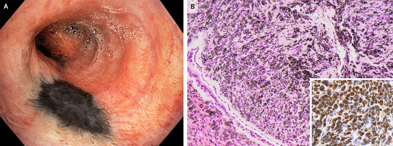
A 77-year-old man undergoing bronchoscopy for evaluation of suspected lung cancer was noted to have black patches on the bronchial mucosa. He had a smoking history of 50 pack-years and a history of pulmonary tuberculosis treated with left upper lobectomy. During bronchoscopy, areas of black discoloration were seen on the mucosa of the left main bronchus and left segmental bronchi (Panel A). A biopsy sample showed ciliated respiratory epithelium with subepithelial accumulation of macrophages containing black pigment (Panel B, hematoxylin and eosin stain). Also noted was the deposition of small, birefringent dust particles of unknown origin, which were thought to possibly be silicates. On immunohistochemical staining, abundant pigment-laden macrophages were identified by their diffuse cytoplasmic positivity for CD68 (Panel B, inset). A diagnosis of bronchial anthracosis — a benign, often incidental finding indicating carbon pigment deposition in the mucosa — was made. Bronchial anthracosis is associated with prolonged exposure to biomass smoke or environmental dust. On further history taking, the patient reported having socialized around open fires for decades of his life in the Middle East, as well as having been exposed to substantial amounts of dust while incarcerated for 10 years. No treatment was recommended for bronchial anthracosis; subsequent management and follow-up focused on the lung cancer that had been confirmed by computed tomography (CT) and CT-guided biopsy.
(2). Dysphagia Lusoria
Ishita Dubeyhttps://orcid.org/0000-0001-9871-3and Neha Wadhwa, Published May 30, 2026,DOI: 10.1056/NEJMicm2600585
Abstract
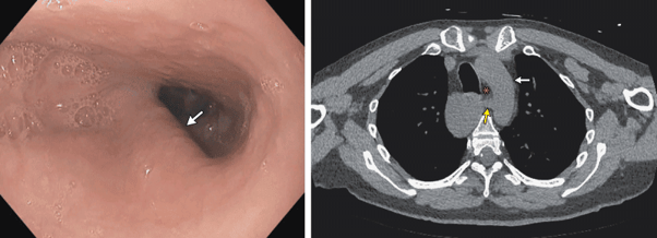
A 44-year-old man with cerebral palsy was referred to the gastroenterology clinic for a 4-year history of choking episodes while eating. Despite treatment for reflux with a proton-pump inhibitor and food-texture modifications to address mild oropharyngeal dysphagia that had been identified in a modified barium-swallow study, the patient’s symptoms had persisted. Physical examination was notable only for long-standing spastic quadriplegia and language delay related to his cerebral palsy. Endoscopic examination revealed luminal narrowing of the upper esophagus by a pulsating, cordlike structure in the posterior wall of the esophagus (Panel A, white arrow, and video). A subsequent computed tomographic scan of the chest showed an aberrant right subclavian artery (Panel B, yellow arrow) originating as a fourth branch of the aortic arch (Panel B, white arrow) and coursing posterior to the esophagus (Panel B, asterisk). A diagnosis of dysphagia lusoria — compression of the esophagus by an aberrant right subclavian artery — was made. Although dysphagia lusoria results from a congenital abnormality, it may not cause symptoms until middle age, when vascular stiffening occurs. Nonsurgical management with continued speech–language therapy for swallowing and dietary modifications was selected by the patient’s health care proxy. Three months after diagnosis, the patient had less frequent choking episodes, continued to eat orally, and maintained his weight.
(3). Chyluria from a Lymphatic–Urinary Fistula
Carles Cañameras, Published June 3, 2026,N Engl J Med 2026;394:2146,DOI: 10.1056/NEJMicm2516914,VOL. 394 NO. 21
Abstract
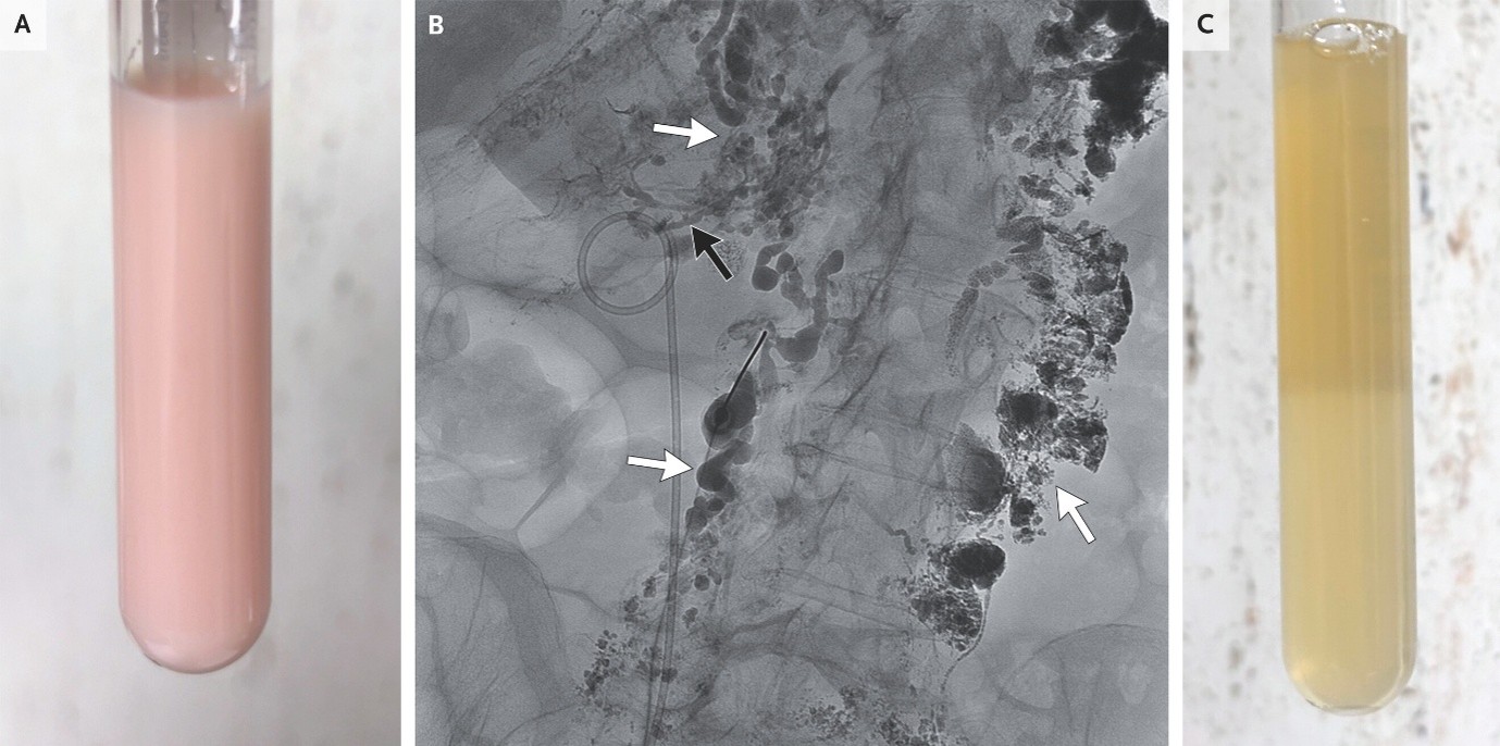
A 56-year-old woman presented to the emergency department with a 3-week history of leg swelling. On physical examination, pitting edema in both legs and a milky appearance to the urine were observed (Panel A). Laboratory studies were notable for a serum albumin level of 18 g per liter (reference range, 35 to 52) and a urinary protein-to-creatinine ratio of 34,958 (with protein measured in milligrams and creatinine measured in grams; reference range, 0 to 200). Further testing of the urine identified elevated levels of chylomicrons and a urinary triglyceride level of 1456 mg per deciliter (reference value, undetectable), findings indicative of chyluria. Chyluria results from abnormal communication between the lymphatic and urinary systems. Possible causes are lymphatic filariasis, congenital lymphatic malformations, post-traumatic lymphatic malformations, and lymphatic compression by tumors. Lymphangiography (after the placement of a right-sided ureteral stent) identified retroperitoneal lymphatic malformations (Panel B, white arrows) with a fistulous communication to the right pyeloureteral junction (Panel B, black arrow). Whether the lymphatic malformations were congenital or acquired was unclear. A diagnosis of chyluria from a lymphatic–urinary fistula was made. The chyluria persisted despite the patient’s following a low-fat diet, so the fistula was subsequently closed by percutaneous transabdominal embolization. At follow-up 3 months later, the appearance of the urine had normalized (Panel C; apparent layering is due to a label on the back of the tube), the patient’s serum albumin levels had improved, and the edema and proteinuria were markedly reduced
(4). Neovascularization of the Disk in Proliferative Diabetic Retinopathy
Ella J. Gehrke et al, Published June 6, 2026,DOI: 10.1056/NEJMicm2601416
Abstract
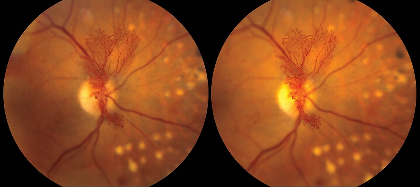
A 58-year-old woman with diabetes complicated by diabetic retinopathy presented for an annual eye examination. Stereo fundus photographs showed neovascularization of the disk extending out from the retinal plane.
Case Presentation
A 58-year-old woman with long-standing type 2 diabetes mellitus complicated by diabetic retinopathy presented to the ophthalmology clinic for an annual eye examination. She had no symptoms affecting her vision. On examination of the right eye, the visual acuity was 20/25. No anteriorsegment neovascularization was seen on slit-lamp examination. Dilated funduscopic examination identified prominent neovascularization of the optic disk, which was documented with stereo fundus photography — an imaging technique in which two images of the retina are obtained at slightly different angles and then viewed through a stereoscopic viewer to create a three-dimensional rendering of retinal structures. This patient’s stereo fundus photographs showed neovascularization of the disk extending out from the plane of the retina into the vitreous cavity. Scars from previous panretinal photocoagulation were also seen (yellow spots in the image background). In the left eye, the visual acuity was 20/25. Dilated fundus examination showed scars from previous panretinal photocoagulation, but no anterior- or posterior-segment neovascularization was visible. A diagnosis of active proliferative diabetic retinopathy in the right eye was made. Prompt treatment to regress the neovascularization in this condition is indicated because the delicate, newly formed vessels may lead to tractional retinal detachment or hemorrhage. Treatment with panretinal photocoagulation and monthly intravitreal anti–vascular endothelial growth factor injections was given. By 3 months after presentation, the patient had near-complete regression of neovascularization of the disk.
(5).Immunosuppression-Associated Peripheral T-Cell Lymphoma
Edward Robert Scheffer Cliff, Published June 13, 2026,N Engl J Med 2026;394:2356
DOI: 10.1056/NEJMicm2501877,VOL. 394 NO. 23
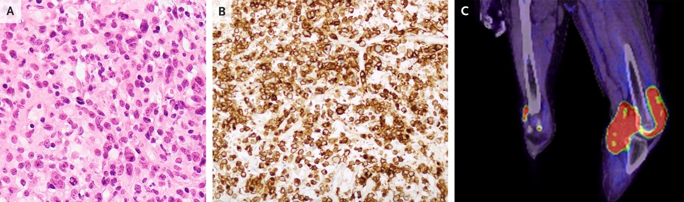
Abstract
A 66-year-old man with a history of Crohn’s disease treated with long-term adalimumab therapy was admitted to the hospital with an 8-week history of fever and swelling of both knees. Physical examination was notable for cachexia and tenosynovitis in both knees (greater in the left knee than in the right). Owing to concern for septic arthritis, a surgical lavage was performed. Histopathological analysis of a biopsy sample of the left knee synovium showed an atypical T-cell infiltrate (Panel A). Immunohistochemical analysis was positive for CD30 (Panel B), CD2, CD3, CD7, CD4, and CD163 and negative for ALK and CD8 and showed 60% expression of Ki-67. Fluorodeoxyglucose positron-emission tomography showed uptake in both knees, with greater uptake in the left knee (Panel C). T-cell gene rearrangement studies showed a clonal T-cell population, and sequencing showed mutations in DNMT3A, TET2, STAT3, JAK1, and TP53. Epstein–Barr encoding region in situ hybridization was negative. A diagnosis of immunosuppression-associated peripheral T-cell lymphoma — a rare condition associated with exposure to thiopurines and tumor necrosis factor inhibitors — was made. Adalimumab therapy was discontinued and multiagent chemotherapy was initiated. At a 14-month follow-up, the lymphoma remained in remission but the Crohn’s disease had flared, leading to treatment with glucocorticoid agents and ustekinumab.
(6).Chromoblastomycosis
Eva R. Parker, Published June 17, 2026,N Engl J Med 2026;394: e54,DOI: 10.1056/NEJMicm2600078, VOL. 394 NO. 23
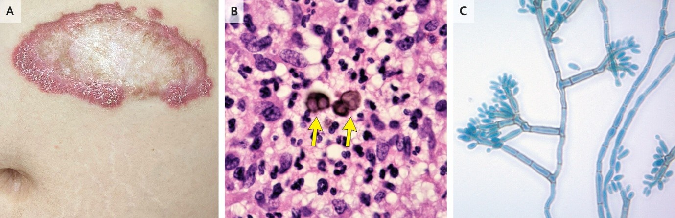
Abstract
A 38-year-old pregnant woman at 30 weeks’ gestation was referred to the dermatology clinic for evaluation of a long-standing, itchy lesion on her abdomen. The lesion had first appeared 9 years earlier after the patient had emigrated from Honduras, where she had worked in agriculture. The lesion had subsequently slowly expanded. On physical examination, a 15-cm erythematous, scaly plaque with a raised border and atrophic center was visible on the left upper abdomen (Panel A). Histopathological analysis of a skin-biopsy sample showed pseudoepitheliomatous hyperplasia with dermal granulomatous infiltrates. Thick-walled, brown, septate spores called muriform cells (Panel B, arrows) were also seen. The presence of such cells (also known as Medlar bodies, sclerotic bodies, or copper pennies) is pathognomonic for chromoblastomycosis. Fungal culture grew a pigmented mold, which showed septate hyphae with loosely branched clavate conidia and prominent denticles on microscopy (Panel C). The causative organism was identified as a fonsecaea species. A diagnosis of chromoblastomycosis with cicatricial morphologic features was confirmed. Chromoblastomycosis is a chronic, progressive implantation mycosis caused by dematiaceous fungi in soil and plant matter. It is typically diagnosed in agricultural workers in tropical or subtropical regions. Postpartum treatment with itraconazole was planned, but the patient was lost to follow-up.
(7). Unheralded Syncope from Ventricular Arrhythmia
Michael Thomas Garcia, Published June 20, 2026,DOI: 10.1056/NEJMicm2600597
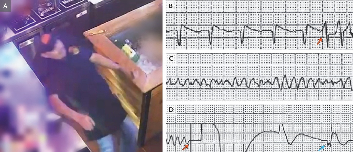
Abstract
A 47-year-old man with ischemic cardiomyopathy, heart failure with a reduced ejection fraction, and an implantable cardioverter–defibrillator (ICD) for the primary prevention of sudden cardiac death presented to the emergency department with a first-time episode of syncope. During the event (which had been captured by video surveillance at work), he collapsed backward without prodromal symptoms and had had a myoclonic jerk (Panel A and video). A few seconds later, he regained consciousness. On presentation, physical examination showed only scalp laceration. Computed tomography of the head showed no intracranial hemorrhage. Interrogation of the ICD identified a premature ventricular contraction during ventricular repolarization — known as the R-on-T phenomenon (Panel B, arrow) — with a normal QT interval; this event had precipitated polymorphic ventricular tachycardia (Panel C). The arrhythmia progressed to ventricular fibrillation, after which a shock was delivered (Panel D, red arrow) and spontaneous electrical activity (blue arrow) returned. A diagnosis of unheralded syncope from ventricular arrhythmia was made. Coronary angiography showed patent stents in the left main and left anterior descending arteries and a known chronic total occlusion of the left circumflex artery. Transthoracic echocardiography showed a stable ejection fraction of 25% with no new wall-motion abnormalities. Treatment with amiodarone was initiated, and beta-blocker therapy was continued. On long-term follow-up, ventricular arrhythmia had not recurred.
(8). Kwashiorkor
Jashir Ahammed Kolathil Gadhiraju MBBS, Manisha Varma, D.N.B., D.C.H. Published June 24, 2026,N Engl J Med 2026;394:2459,DOI: 10.1056/NEJMicm2602658, VOL. 394 NO. 24
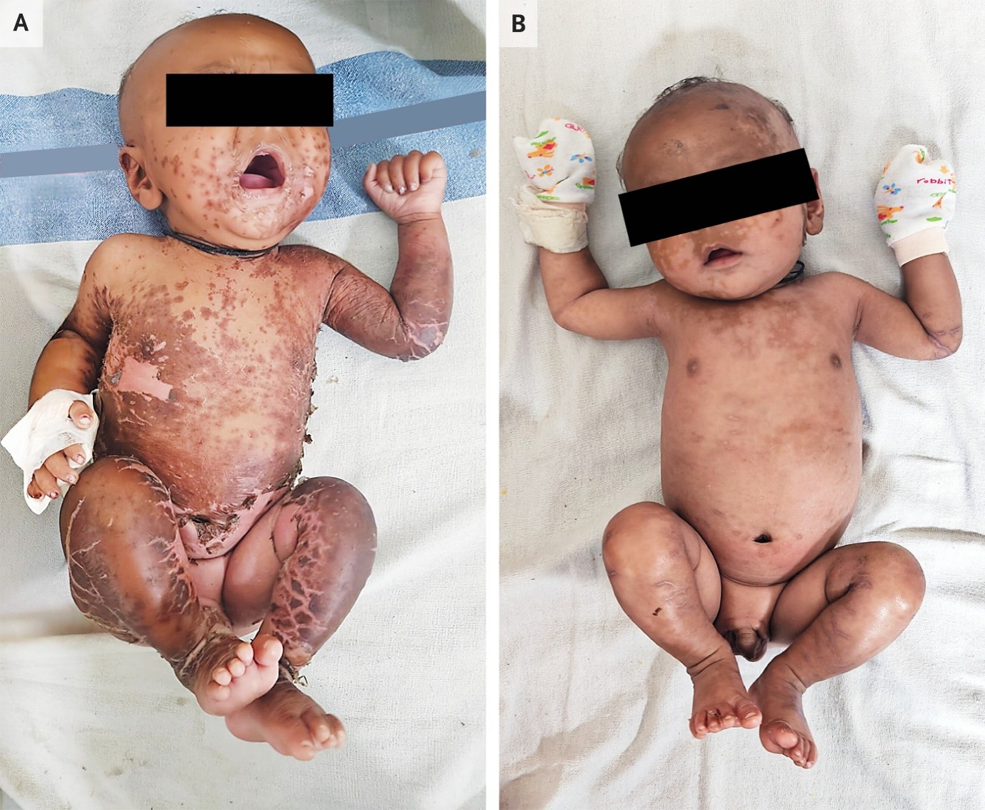
Abstract
A 5-month-old full-term baby boy was brought to the outpatient clinic with a 3-week history of a rash. He had consumed breast milk exclusively until 20 days before presentation, when he had been switched to diluted formula owing to his mother’s ongoing challenges with limited breast-milk supply. His weight-for-age z score was −3.4, and his length-for-age z score was −5.6. On physical examination, the baby was irritable, with sparse, wispy, hypopigmented hair and pitting edema of the arms and legs. Desquamation of hyperpigmented patches was seen on the legs, arms, and trunk; the underlying skin was hypopigmented (Panel A). A serum albumin level was 1.9 g per deciliter (reference range, 3.5 to 5.0). A diagnosis of kwashiorkor — also known as edematous malnutrition or severe acute malnutrition with pitting edema of the arms and legs — was made.
Kwashiorkor may be diagnosed in malnourished children on the basis of the presence of pitting edema, regardless of height and weight values. The typical dermatosis, as seen in this baby, resembles flaking paint. Inpatient treatment with a therapeutic milk formula (F-75) was initiated. On day 7 of the admission, the baby was more active, resolving skin lesions (Panel B). At a follow-up visit 2 weeks after discharge, the rash had abated and weight gain had normalized.
(9).Cat Scratch Disease
Reena D. Wadhwa, Published June 27, 2026,DOI: 10.1056/NEJMicm2602360
Abstract
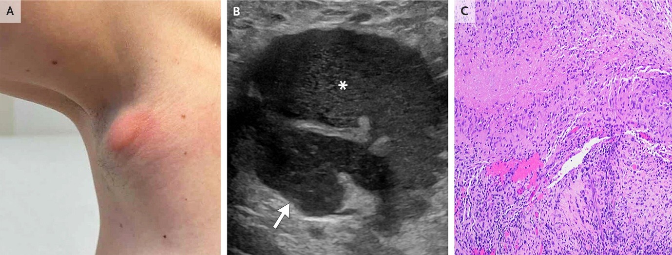
A previously healthy 40-year-old woman presented to the primary care clinic with a 1-month history of fevers and a 3-week history of swelling in her right armpit that had not abated with a course of trimethoprim–sulfamethoxazole. On physical examination, there was tender lymphadenopathy with overlying erythema in the right axilla (Panel A, anterior view). A cluster of papules and a scar were noted on the distal right index finger, where the patient recalled being bitten by a stray kitten 2.5 months before presentation. Owing to concern for cat scratch disease, azithromycin was prescribed. The fevers abated but the lymphadenopathy persisted; therefore, further evaluation was performed. Ultrasonography showed enlarged lymph nodes (Panel B, arrow) with an adjacent fluid collection (Panel B, asterisk). Histopathological analysis of a core-needle biopsy specimen obtained from a lymph node showed palisading histiocytes surrounding caseous necrosis and granulomatous inflammation (Panel C). Polymerase-chain-reaction testing of a tissue specimen was positive for Bartonella henselae. IgG serologic testing for bartonella was also positive. A diagnosis of cat scratch disease was confirmed. Treatment with doxycycline and rifampin was given. Three weeks after initiation of treatment, the lymphadenopathy had worsened and the lymph nodes were leaking pus, so débridement was performed. Three months after initiation of treatment, the symptoms resolved.
(10). Trampoline Fracture
Tushar Kumar, Published July 1, 2026,N Engl J Med 2026;395: e1. DOI: 10.1056/NEJMicm2601744,VOL. 395 NO. 1
Abstract
A previously healthy 4-year-old boy presented to the emergency department with a 1-hour history of leg pain and an inability to bear weight on his left side. While jumping on a trampoline with his father at home, he had leaped higher than usual, landed on his feet, and then felt the leg pain. Physical examination was notable for knee swelling and tenderness to palpation on the left side. Radiographs of the left tibia and fibula showed a nondisplaced fracture of the proximal tibial metaphysis (Panel A, frontal view, white arrow) extending up to the growth plate (black arrow). A diagnosis of a trampoline fracture was made.
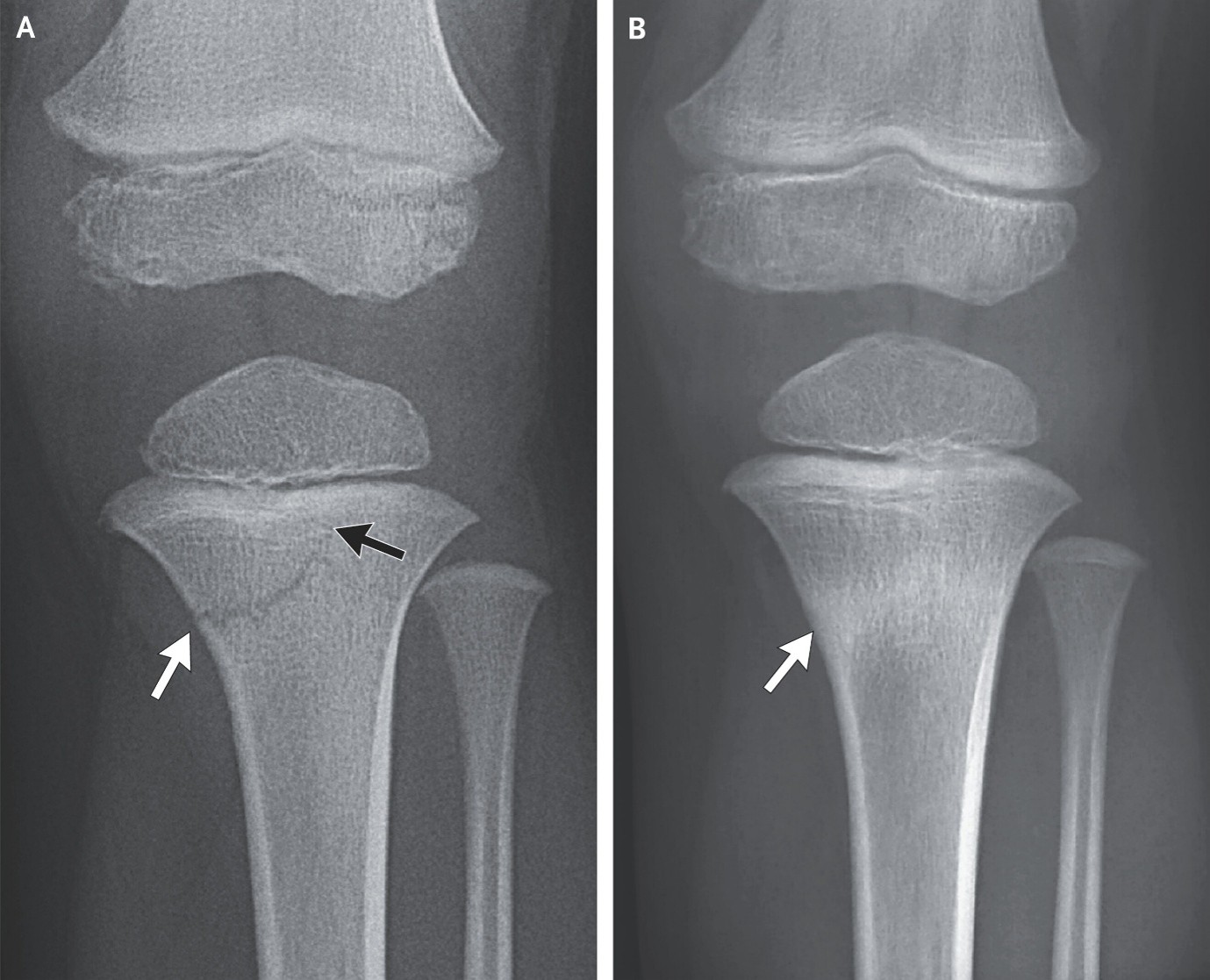
Trampoline fractures in children typically manifest as transverse, proximal tibial fractures owing to compressive axial forces on the bone while bouncing. The risk of these injuries increases when a heavier person is also jumping and generating increased recoil of the trampoline. Growth-plate involvement in these fractures may result in altered limb growth. After 4 weeks in a cast with no weight bearing, the child started to resume normal activities. Six months after the injury, a repeat radiograph showed interval healing with mild residual sclerosis and no complications of growth-plate involvement (Panel B, arrow).
(11). Condyloma Acuminata of the Urethra
Jung Hoon Bae, ,Published July 8, 2026,N Engl J Med 2026;395:175,DOI: 10.1056/NEJMicm2603369.VOL. 395 NO. 2
Abstract
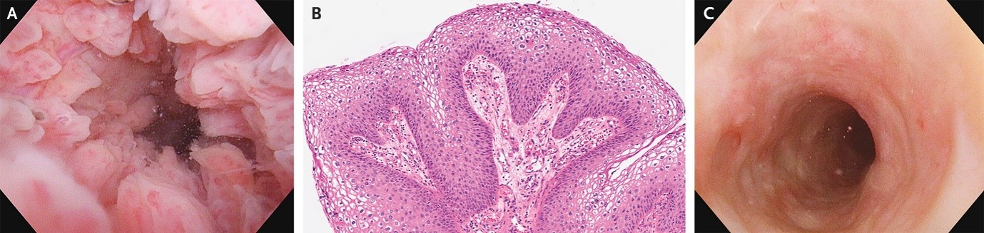
A 42-year-old man presented to the urology clinic with a 2-week history of urinary frequency and weak stream. Four months before presentation, the patient had undergone stem-cell transplantation for acute lymphoblastic leukemia, which had been complicated by chronic graft-versus-host disease that had led to prolonged immunosuppressive therapy. One month before presentation, the patient had noted multiple small bumps on his penis. He was in a monogamous relationship with one sexual partner. Physical examination was notable for several small papules on the penile shaft. Urinalysis showed 9 white cells per high-power field, and a urine culture was negative. Testing for human immunodeficiency virus was also negative. Urethroscopy showed papillomatous lesions carpeting the urethral mucosa (Panel A). The penile papules were excised and identified on histopathological analysis as condyloma acuminata (genital warts), and resected urethral tissue showed papillomatous squamous proliferation with koilocytic atypia (Panel B, hematoxylin and eosin stain). A polymerase-chain-reaction assay of the excised penile-lesion specimen was positive for human papilloma virus (HPV) type 11 (the patient had not been vaccinated against HPV). A diagnosis of condyloma acuminata of the urethra was made. Treatment consisted of transurethral ablation of the lesions followed by intraurethral instillations of 5-fluorouracil and additional transurethral ablations. At follow-up 11 months after the initial diagnosis, the urethral lesions had not recurred (Panel C).
(12). Descending Thoracic Aortic Aneurysm
Maithili Charan Gattu, M.B., B.S., and Mehul K, M.B., B.S., M.D. Published July 11, 2026,DOI: 10.1056/NEJMicm2505469
Abstract
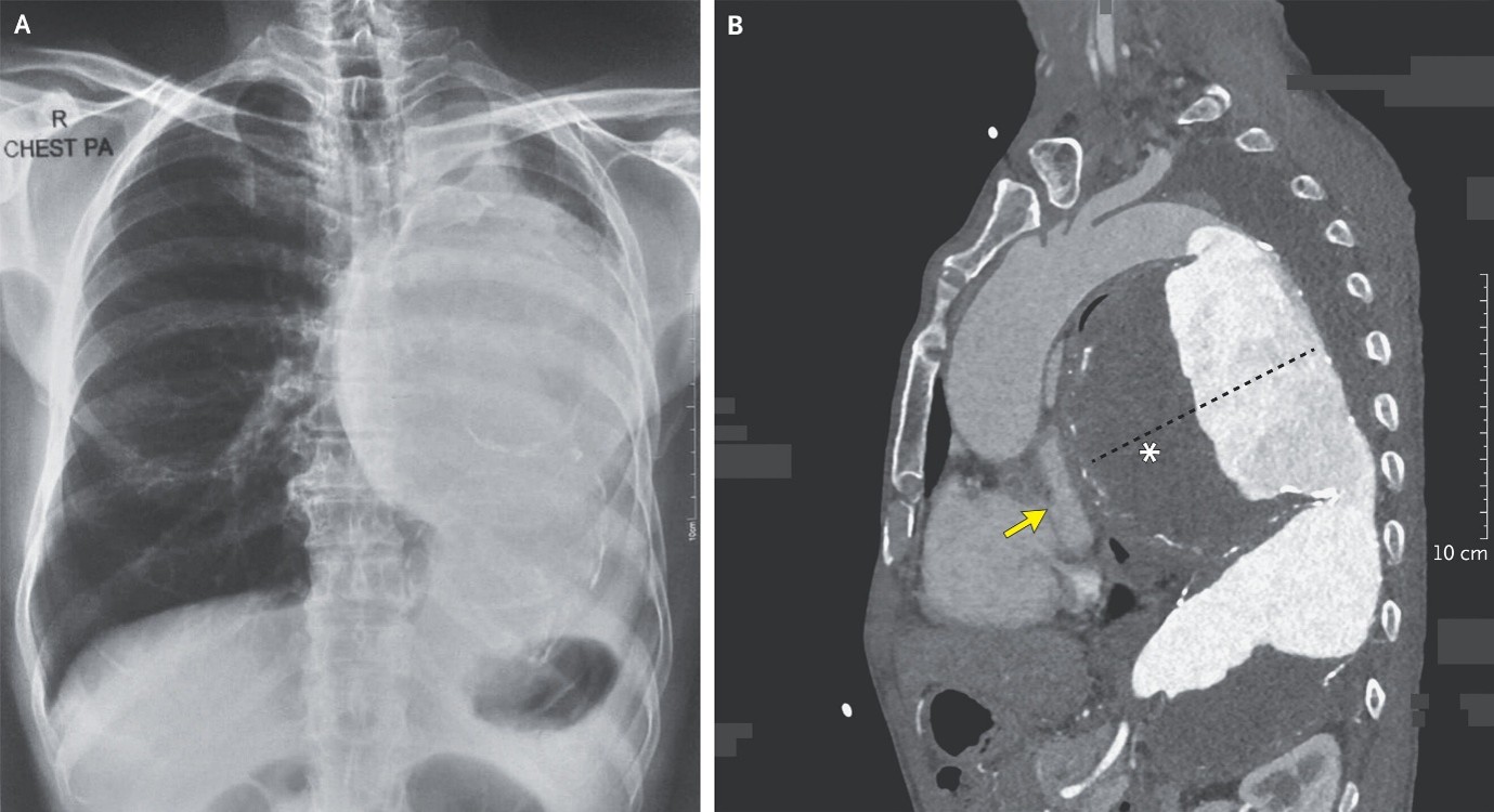
A 54-year-old man with no known medical history presented to the emergency department with chest heaviness that had persisted for 2 months and a 2-week history of shortness of breath. His heart rate was 93 beats per minute, and his blood pressure was 140/93 mm Hg. On physical examination, there were visible pulsations in the fourth through seventh intercostal spaces on the left side (Video 1A). Elevated jugular venous pressure and decreased breath sound on the left side of the chest were noted. A radiograph of the chest showed a large, rounded opacity occupying the left hemithorax (Panel A). Point-of-care ultrasonography of the heart showed a rounded structure with both anechoic and echogenic components that compressed the left atrium (Video 1B, asterisk). Computed tomographic angiography of the chest showed a large, fusiform aneurysm in the descending thoracic aorta (Panel B, dashed line; sagittal view). The aneurysm contained an eccentric thrombus (asterisk) and compressed the left atrium (arrow). Emergency open surgical repair of the aneurysm was performed. However, the patient died on postoperative day 3. An autopsy was not performed. The histopathological report was recorded on paper and was not available for retrospective review; therefore, the cause of the aneurysm is unknown.