
Disseminated tuberculosis presenting as multiple cerebral ring-enhancing lesions mimicking neurocysticercosis in a diabetic male with fever of unknown origin: A case report and review of literature
Gowdham Pannirselvam*
Senior Nurse Educator, Kauvery Hospital, Marathahalli, Bangalore
*Correspondence
Abstract
Background
Disseminated tuberculosis is a severe form of Mycobacterium tuberculosis infection resulting from lymphohematogenous spread involving two or more non-contiguous organs. Central nervous system (CNS) tuberculosis represents one of the most devastating manifestations of extrapulmonary tuberculosis and frequently poses diagnostic challenges due to its varied clinical and radiological presentations. Multiple cerebral tuberculomas may closely mimic neurocysticercosis, metastatic lesions, fungal infections, or cerebral abscesses, often leading to delays in diagnosis and treatment.
Case Presentation
A 39-year-old male with known type 2 diabetes mellitus presented with intermittent high-grade fever of 15 days duration and significant weight loss. Initial investigations performed elsewhere for common infectious causes including dengue, malaria, typhoid fever, and bacterial infections were negative. Subsequently, the patient developed headache, diplopia, blurring of vision, vertigo, vomiting, focal neurological deficits, and an episode of collapse. Magnetic resonance imaging of the brain demonstrated multiple scattered ring-enhancing lesions with surrounding edema. Initial radiological differentials included neurocysticercosis and cerebral tuberculomas. Cerebrospinal fluid analysis revealed lymphocytic pleocytosis, elevated protein, and low glucose levels. High-resolution computed tomography of the chest showed bilateral randomly distributed micronodular opacities suggestive of miliary tuberculosis, while PET-CT demonstrated supraclavicular and mediastinal lymphadenopathy. Based on the combined clinical, radiological, and laboratory findings, a diagnosis of disseminated tuberculosis with cerebral tuberculomas and probable tuberculous meningitis was established. The patient was managed with anti-tubercular therapy, corticosteroids, electrolyte correction, and supportive care, resulting in gradual clinical improvement.
Conclusion
This case highlights the importance of considering disseminated tuberculosis in patients presenting with fever of unknown origin and multiple ring-enhancing cerebral lesions, particularly in endemic regions. Early multidisciplinary evaluation and correlation of systemic and neuroimaging findings are essential to avoid diagnostic delays and improve outcomes.
Keywords: Disseminated tuberculosis; Cerebral tuberculoma; Neurocysticercosis; Tuberculous meningitis; Miliary tuberculosis; Ring-enhancing lesions; Fever of unknown origin.
Introduction
Tuberculosis (TB) remains one of the leading infectious causes of morbidity and mortality worldwide despite significant advances in diagnostic and therapeutic strategies. According to the World Health Organization, tuberculosis continues to affect millions of individuals annually, particularly in low- and middle-income countries. Although pulmonary disease remains the most common manifestation, extrapulmonary tuberculosis accounts for approximately 15–20% of reported cases and may involve virtually any organ system. Among these manifestations, central nervous system tuberculosis represents one of the most severe forms because of its high rates of neurological disability and mortality if diagnosis and treatment are delayed. [1,2]
Disseminated tuberculosis occurs when Mycobacterium tuberculosis spreads through hematogenous or lymphatic routes from a primary focus of infection to distant organs. Commonly affected sites include the lungs, lymph nodes, meninges, liver, spleen, bones, and brain. Risk factors for dissemination include diabetes mellitus, HIV infection, malnutrition, immunosuppressive therapy, malignancy, and chronic systemic illnesses. Diabetes mellitus has emerged as an important contributor to both pulmonary and extrapulmonary tuberculosis owing to impaired cellular immune responses and altered macrophage function. [3,4]
Anatomy of the involved structures
The central nervous system comprises the brain, meninges, ventricular system, and spinal cord. The cerebral hemispheres receive blood supply through branches of the internal carotid and vertebrobasilar systems. The meninges consist of three layers: dura mater, arachnoid mater, and pia mater. The subarachnoid space between the arachnoid and pia contains cerebrospinal fluid (CSF), which circulates through the ventricular system and provides metabolic and mechanical support to the brain. Tuberculous infection may involve both the meninges and brain parenchyma, resulting in tuberculous meningitis and cerebral tuberculomas. [5]
Tuberculomas are localized granulomatous masses composed of epithelioid cells, Langhans giant cells, lymphocytes, and central caseous necrosis. They may develop anywhere within the cerebral hemispheres, cerebellum, brainstem, or basal ganglia. Depending on their size and location, tuberculomas may produce focal neurological deficits, seizures, raised intracranial pressure, visual disturbances, or cognitive impairment. [6]
The lungs serve as the primary portal of entry for Mycobacterium tuberculosis. In miliary tuberculosis, hematogenous dissemination results in numerous tiny granulomatous lesions distributed throughout both lungs, producing the characteristic millet-seed appearance on imaging studies. Disseminated infection may further involve lymphatic structures including supraclavicular, mediastinal, and abdominal lymph nodes, as observed in the present case. [7]
Pathophysiology
Tuberculosis begins following inhalation of aerosolized droplets containing Mycobacterium tuberculosis. After reaching the alveoli, bacilli are phagocytosed by alveolar macrophages. In susceptible individuals, organisms evade intracellular destruction and proliferate within macrophages. Subsequent dissemination through lymphatic and hematogenous pathways may occur before adequate cellular immunity develops. [8]
Central nervous system tuberculosis is believed to result from hematogenous seeding of bacilli into the meninges or brain parenchyma, forming microscopic subpial or subependymal lesions known as Rich foci. Rupture of these lesions into the subarachnoid space can result in tuberculous meningitis, whereas progressive enlargement within the brain parenchyma produces tuberculomas. [9]
Radiologically, cerebral tuberculomas often appear as ring-enhancing lesions due to peripheral granulomatous inflammation surrounding a necrotic center. These findings frequently overlap with neurocysticercosis, pyogenic abscesses, fungal infections, metastases, and primary brain tumors. Consequently, diagnosis requires careful integration of clinical presentation, imaging findings, cerebrospinal fluid analysis, and evidence of systemic tuberculosis. [10]
The present report describes a diagnostically challenging case of disseminated tuberculosis presenting initially as fever of unknown origin and subsequently manifesting as multiple cerebral ring-enhancing lesions mimicking neurocysticercosis, highlighting the importance of multidisciplinary evaluation and timely recognition of this potentially life-threatening condition.
Case presentation
A 39-year-old male with a known history of type 2 diabetes mellitus presented with intermittent high-grade fever for 15 days associated with generalized weakness, loss of appetite, and significant weight loss of approximately 8 kg. The patient initially sought medical attention at another healthcare facility where extensive investigations were performed to identify the etiology of fever. Screening for common tropical and bacterial infections, including dengue fever, malaria, typhoid fever, and routine blood cultures, was reportedly negative. Despite symptomatic treatment and empirical therapy, the fever persisted.
One day before admission to our institution, the patient developed acute onset severe headache associated with room-spinning sensation, blurring of vision, diplopia, recurrent vomiting, and focal neurological deficits. Subsequently, he experienced a collapse episode at home, prompting emergency medical evaluation. There was no history of seizures, loss of consciousness, recent travel, exposure to tuberculosis, or prior neurological illness. HIV risk factors were absent, and HIV serology was later confirmed to be negative.
On presentation, the patient was conscious, alert, and oriented. Vital signs were stable. General examination demonstrated significant constitutional symptoms including weight loss and fatigue. Neurological examination revealed diplopia and mild focal neurological weakness. Meningeal signs were not prominent at presentation. Cardiovascular and abdominal examinations were unremarkable.
Given the acute neurological symptoms, urgent neuroimaging was performed to exclude cerebrovascular events, intracranial haemorrhage, and space-occupying lesions.
Investigations
Laboratory Investigations
Initial hematological and biochemical investigations demonstrated evidence of systemic inflammation. Evaluation for common infectious etiologies including dengue fever, malaria, enteric fever, and routine bacterial infections was negative. HIV serology was non-reactive.
Neuroimaging
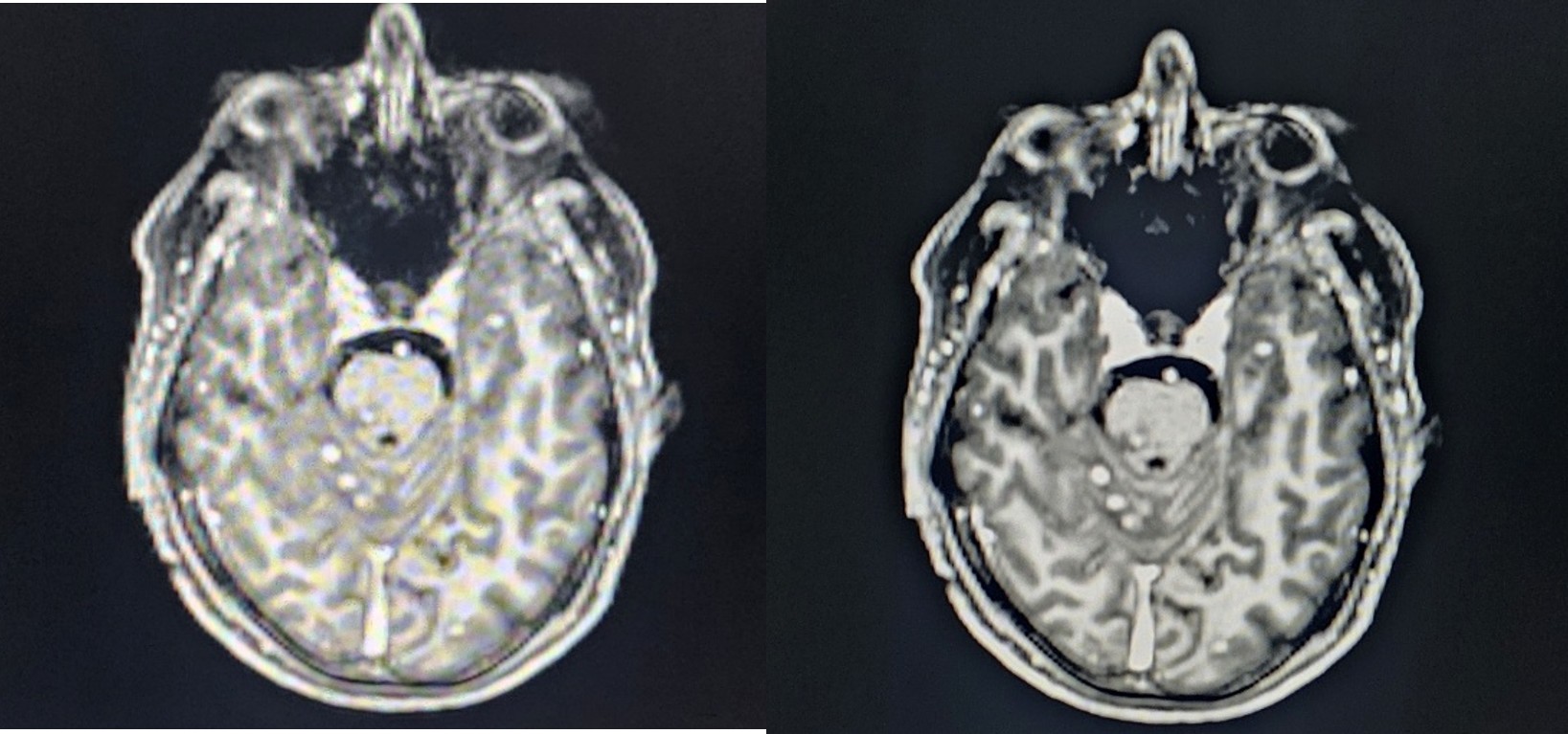
Magnetic resonance imaging (MRI) of the brain with contrast revealed multiple scattered ring-enhancing lesions involving both cerebral hemispheres. Several lesions demonstrated surrounding vasogenic edema. The radiological appearance raised suspicion for infective granulomatous pathology.
The principal radiological differentials included:
- Neurocysticercosis
- Cerebral tuberculomas
- Fungal granulomas
- Metastatic lesions
- Pyogenic abscesses
The multiplicity of lesions and their ring-enhancing appearance initially created significant diagnostic uncertainty. In endemic regions, both neurocysticercosis and cerebral tuberculomas represent important causes of multiple ring-enhancing lesions and frequently mimic one another on imaging studies. [11,12]
Cerebrospinal Fluid Analysis
Lumbar puncture was subsequently performed for further evaluation. Cerebrospinal fluid examination demonstrated:
- Lymphocytic pleocytosis
- Elevated protein concentration
- Reduced glucose levels
- Total cell counts approximately 435 cells/mm³
GeneXpert testing for Mycobacterium tuberculosis was negative.
Despite the negative molecular test, the biochemical profile remained strongly suggestive of tuberculous meningitis. The sensitivity of CSF GeneXpert may be limited in paucibacillary disease and therefore does not exclude CNS tuberculosis. [13]
Chest Imaging
High-resolution computed tomography (HRCT) of the chest revealed:
- Bilateral randomly distributed micronodular opacities
- Diffuse miliary pattern
- Bibasal subsegmental atelectatic changes
- Trace pleural effusion
These findings significantly shifted diagnostic consideration toward disseminated tuberculosis with pulmonary involvement.
PET-CT Findings
PET-CT demonstrated metabolically active lymphadenopathy involving:
- Left supraclavicular lymph nodes
- Retrosternal lymph nodes
The combination of miliary pulmonary nodules, systemic lymphadenopathy, constitutional symptoms, and CNS lesions strongly favored disseminated tuberculosis.
Additional Clinical Findings
During hospitalization, serial biochemical investigations demonstrated:
- Hyponatremia
- Elevated liver enzymes
- Episodes of recurrent vomiting
Hyponatremia was attributed to syndrome of inappropriate antidiuretic hormone secretion (SIADH), a recognized complication of CNS tuberculosis.
Differential diagnosis
The diagnosis of multiple ring-enhancing lesions remains one of the most challenging scenarios encountered in clinical neurology and neuroradiology. Several conditions were considered during the evaluation of this patient.
Neurocysticercosis: Neurocysticercosis was initially considered because it represents one of the most common causes of multiple ring-enhancing lesions in endemic regions. The disease results from CNS infection by the larval stage of Taenia solium. Neuroimaging frequently demonstrates multiple cystic or ring-enhancing lesions with surrounding edema. However, the presence of prolonged fever, marked weight loss, miliary pulmonary nodules, lymphadenopathy, and CSF findings suggestive of meningitis were less characteristic of neurocysticercosis and favoured tuberculosis. [14]
Cerebral Tuberculoma: Cerebral tuberculoma became the leading diagnosis because of the coexistence of constitutional symptoms, pulmonary abnormalities, lymphadenopathy, and CSF findings suggestive of CNS tuberculosis. Tuberculomas commonly appear as ring-enhancing lesions and may occur with or without concomitant tuberculous meningitis. [15]
Metastatic Disease: Multiple cerebral metastases were considered because metastatic lesions frequently appear as multiple ring-enhancing lesions at the gray-white matter junction. However, the patient’s age, clinical history, absence of a known primary malignancy, and associated infectious findings made metastatic disease less likely. [16]
Pyogenic Brain Abscess: Multiple cerebral abscesses can produce ring-enhancing lesions and neurological deficits. However, the absence of bacteraemia, negative cultures, lack of significant leukocytosis, and associated pulmonary findings reduced the likelihood of this diagnosis. [17]
Fungal Infection: Disseminated fungal infections such as cryptococcosis or aspergillosis may produce multiple cerebral lesions, particularly in immunocompromised hosts. Negative HIV status and the characteristic pulmonary findings favored tuberculosis over fungal disease. [18]
Final diagnosis
Based on the integration of clinical, radiological, laboratory, and systemic findings, a final diagnosis of disseminated tuberculosis was established, comprising:
- Multiple cerebral tuberculomas
- Probable tuberculous meningitis
- Miliary pulmonary tuberculosis
- Mediastinal and supraclavicular lymphadenopathy
- SIADH-associated hyponatremia
- Type 2 diabetes mellitus
Treatment
The patient was managed through a multidisciplinary team consisting of neurologists, pulmonologists, infectious disease specialists, ophthalmologists, and critical care physicians. Anti-tubercular therapy (ATT) was initiated based on strong clinicoradiological evidence of disseminated tuberculosis despite the absence of microbiological confirmation. Standard first-line therapy consisting of isoniazid, rifampicin, pyrazinamide, and ethambutol was commenced according to national treatment guidelines. [19]. Adjunctive corticosteroid therapy was administered because of probable CNS tuberculosis and cerebral edema. Corticosteroids have been shown to reduce mortality and neurological complications in tuberculous meningitis. [20]
Supportive treatment included:
- Intravenous fluids
- Electrolyte correction
- Glycemic control
- Antiemetics
- Nutritional supplementation
- Physiotherapy and rehabilitation
During hospitalization, the patient developed elevated liver enzymes and recurrent vomiting suggestive of drug-induced hepatotoxicity. Consequently, ATT was temporarily withheld for four days while serial liver function tests were monitored. Following improvement in hepatic parameters, anti-tubercular therapy was gradually reintroduced. Hyponatremia secondary to SIADH was managed through fluid restriction, electrolyte monitoring, and corrective therapy. Progressive normalization of serum sodium levels was observed.
Rationale for choice of treatment
Alternative treatment approaches were considered during evaluation.
Surgical intervention was not indicated because the cerebral lesions were multiple, deep-seated, and lacked evidence of significant mass effect requiring decompression. Biopsy was considered but deferred due to the convincing clinic-radiological diagnosis and favourable response to medical treatment. Therefore, anti-tubercular therapy combined with corticosteroids was considered the most appropriate management strategy, addressing both systemic tuberculosis and CNS involvement while avoiding unnecessary invasive procedures.
Outcome and follow-up
The patient demonstrated gradual clinical improvement following initiation of anti-tubercular therapy, corticosteroids, and supportive management. Fever spikes progressively subsided, appetite improved, and the patient reported reduction in headache and visual symptoms. Neurological examination showed stabilization of focal deficits without development of new neurological manifestations. No seizure episodes occurred during hospitalization.
The patient’s hospital course was complicated by transient drug-induced hepatotoxicity and hyponatremia secondary to syndrome of inappropriate antidiuretic hormone secretion (SIADH). Both complications were recognized early and managed appropriately. Following temporary interruption and subsequent reintroduction of anti-tubercular therapy, liver function parameters improved. Serial monitoring demonstrated correction of serum sodium levels and improvement in overall clinical status.
At discharge, the patient was afebrile, hemodynamically stable, and neurologically improved. He was advised to continue anti-tubercular therapy under close supervision, maintain strict glycemic control, undergo periodic liver function monitoring, and attend regular follow-up visits with Neurology, Pulmonology, and Infectious Disease specialists. Repeat neuroimaging and chest imaging were planned to assess treatment response and radiological resolution of lesions.
Discussion
Tuberculosis remains a major global health concern, particularly in developing countries where disease burden continues to be substantial. Although pulmonary tuberculosis is the most frequently encountered form, extrapulmonary manifestations account for a significant proportion of cases and often pose diagnostic challenges because of their diverse clinical presentations. Central nervous system tuberculosis is among the most severe forms of extrapulmonary disease, contributing significantly to morbidity and mortality. Delayed diagnosis may result in irreversible neurological deficits, hydrocephalus, seizures, stroke, and death. [1,2]
The present case highlights several important clinical and diagnostic lessons. First, the patient initially presented with prolonged fever without an obvious source, fulfilling the criteria for fever of unknown origin (FUO). Fever of unknown origin remains a challenging clinical entity requiring systematic evaluation. In countries where tuberculosis is endemic, extrapulmonary and disseminated tuberculosis should always remain an important differential diagnosis in patients with persistent fever and constitutional symptoms, even when initial microbiological investigations are negative. [3]
An interesting feature of this case was the evolution of neurological symptoms leading to neuroimaging. The presence of headache, diplopia, blurring of vision, vomiting, and focal neurological deficits prompted urgent MRI evaluation. Neuroimaging demonstrated multiple ring-enhancing lesions, creating a broad differential diagnosis. Ring-enhancing lesions represent a common neuroradiological challenge and may be encountered in infections, neoplasms, inflammatory disorders, and vascular lesions. The differential diagnosis commonly includes neurocysticercosis, cerebral tuberculoma, pyogenic abscess, toxoplasmosis, fungal infections, primary CNS lymphoma, and metastatic disease. [4,5]
The distinction between neurocysticercosis and cerebral tuberculoma is particularly important in endemic regions because both conditions frequently present as multiple ring-enhancing lesions. Neurocysticercosis results from infection with the larval form of Taenia solium and remains one of the most common parasitic infections affecting the central nervous system. In contrast, cerebral tuberculomas arise from hematogenous dissemination of Mycobacterium tuberculosis into the brain parenchyma. Both diseases may demonstrate similar radiological appearances, making differentiation challenging based solely on imaging. Clinical history, systemic findings, cerebrospinal fluid analysis, and evidence of extracranial disease often provide essential clues for diagnosis. [6,7]
In the present case, the radiological differential initially included neurocysticercosis. However, several findings favoured tuberculosis. The patient had prolonged fever, significant weight loss, lymphocytic cerebrospinal fluid pleocytosis, low CSF glucose levels, elevated protein concentration, miliary pulmonary nodules, and systemic lymphadenopathy. These features collectively supported a diagnosis of disseminated tuberculosis rather than isolated parasitic infection. This emphasizes the importance of integrating radiological findings with clinical and laboratory data rather than relying on imaging alone. [8]
The pathogenesis of CNS tuberculosis involves hematogenous dissemination of tubercle bacilli from a primary focus, usually within the lungs. Bacilli may seed the meninges or brain parenchyma and form microscopic lesions known as Rich foci. Subsequent rupture of these foci into the subarachnoid space leads to tuberculous meningitis, whereas progressive enlargement within the brain parenchyma results in tuberculoma formation. The coexistence of cerebral tuberculomas and CSF findings suggestive of meningitis in this patient supports this well-recognized pathogenic mechanism. [9]
An additional noteworthy aspect of this case was the negative GeneXpert result despite overwhelming clinical evidence of tuberculosis. Molecular diagnostic tests have significantly improved the diagnosis of tuberculosis; however, their sensitivity may be limited in paucibacillary specimens such as cerebrospinal fluid. Therefore, a negative GeneXpert result should not exclude CNS tuberculosis when clinical suspicion remains high. Current guidelines emphasize the importance of clinicoradiological diagnosis in situations where microbiological confirmation cannot be achieved. [10]
The presence of miliary pulmonary tuberculosis was a critical clue that guided the diagnostic process. Miliary tuberculosis results from widespread hematogenous dissemination of Mycobacterium tuberculosis and is characterized radiologically by numerous small pulmonary nodules distributed throughout both lungs. CNS involvement occurs in a significant proportion of patients with miliary tuberculosis and is associated with poorer outcomes. Early recognition and treatment are therefore essential. [11]
Diabetes mellitus likely contributed to disease progression in this patient. Numerous studies have demonstrated that diabetes increases susceptibility to tuberculosis and adversely affects disease outcomes. Impaired macrophage function, reduced cellular immunity, and chronic inflammatory dysregulation contribute to increased risk of both pulmonary and extrapulmonary disease. Patients with diabetes are also more likely to develop severe forms of tuberculosis, delayed treatment response, and treatment-related complications. [12]
Treatment of cerebral tuberculomas primarily involves prolonged anti-tubercular therapy. Surgical intervention is generally reserved for patients with diagnostic uncertainty, significant mass effect, obstructive hydrocephalus, or failure of medical therapy. In the present case, medical management was selected because the lesions were multiple, imaging findings were consistent with granulomatous disease, and systemic evidence strongly supported tuberculosis. Furthermore, the patient demonstrated favourable clinical response following initiation of therapy, confirming the appropriateness of the chosen treatment strategy. [13]
Adjunctive corticosteroid therapy remains an important component of management in CNS tuberculosis. Randomized controlled trials have demonstrated reductions in mortality and neurological complications among patients receiving corticosteroids in addition to anti-tubercular therapy. Corticosteroids reduce inflammation, cerebral edema, and intracranial pressure, thereby improving neurological outcomes. [14]
The occurrence of SIADH-associated hyponatremia in this patient is also consistent with previous reports of CNS tuberculosis. Hyponatremia may result from SIADH or cerebral salt wasting syndrome and is associated with increased morbidity if not promptly recognized and treated. Regular electrolyte monitoring is therefore essential during the management of CNS tuberculosis. [15]
Overall, this case underscores the importance of maintaining a high index of suspicion for disseminated tuberculosis in patients presenting with prolonged fever, constitutional symptoms, and multiple ring-enhancing brain lesions. Early multidisciplinary involvement, careful interpretation of imaging findings, and consideration of systemic disease manifestations were crucial in establishing the diagnosis and initiating timely treatment.
Conclusion
Disseminated tuberculosis remains a diagnostic challenge because of its ability to mimic a wide range of infectious, inflammatory, and neoplastic disorders. This case illustrates an unusual presentation of disseminated tuberculosis manifesting as fever of unknown origin and multiple cerebral ring-enhancing lesions initially suspected to represent neurocysticercosis. The coexistence of miliary pulmonary disease, lymphocytic cerebrospinal fluid findings, systemic lymphadenopathy, and constitutional symptoms ultimately led to the correct diagnosis. Early recognition and initiation of anti-tubercular therapy resulted in favourable clinical outcomes. Clinicians practicing in tuberculosis-endemic regions should maintain a high level of suspicion for CNS tuberculosis when evaluating patients with unexplained neurological symptoms and ring-enhancing brain lesions.
Learning points
- Disseminated tuberculosis should be considered in patients presenting with fever of unknown origin and constitutional symptoms.
- Multiple ring-enhancing cerebral lesions require careful evaluation because they may mimic neurocysticercosis, metastases, abscesses, or fungal infections.
- A negative GeneXpert result does not exclude CNS tuberculosis.
- Miliary pulmonary tuberculosis may provide an important clue to underlying disseminated disease.
- Diabetes mellitus is an important risk factor for severe and extrapulmonary tuberculosis.
- Multidisciplinary collaboration is often essential for establishing the diagnosis of complex tuberculosis cases.
- Early initiation of anti-tubercular therapy can significantly improve neurological and systemic outcomes.
References
- World Health Organization. Global tuberculosis report 2024. Geneva: World Health Organization; 2024.
- Pai M, Behr MA, Dowdy D, Dheda K, Divangahi M, Boehme CC, et al. Tuberculosis. Nat Rev Dis Primers. 2016;2:16076.
- Golden MP, Vikram HR. Extrapulmonary tuberculosis: an overview. Am Fam Physician. 2005;72(9):1761-1768.
- Donnelley MA, Zhu X, Akinbi HT. Tuberculosis and diabetes mellitus. Clin Chest Med. 2019;40(4):739-751.
- Standring S, editor. Gray’s Anatomy: The Anatomical Basis of Clinical Practice. 42nd ed. London: Elsevier; 2020.
- Rock RB, Olin M, Baker CA, Molitor TW, Peterson PK. Central nervous system tuberculosis: pathogenesis and clinical aspects. Clin Microbiol Rev. 2008;21(2):243-261.
- Sharma SK, Mohan A, Sharma A, Mitra DK. Miliary tuberculosis: new insights into an old disease. Lancet Infect Dis. 2005;5(7):415-430.
- Davis AG, Rohlwink UK, Proust A, Figaji AA, Wilkinson RJ. The pathogenesis of tuberculous meningitis. J Leukoc Biol. 2019;105(2):267-280.
- Wilkinson RJ, Rohlwink U, Misra UK, Van Crevel R, Mai NTH, Dooley KE, et al. Tuberculous meningitis. Nat Rev Neurol. 2017;13(10):581-598.
- Bernaerts A, Vanhoenacker FM, Parizel PM, Van Goethem JW, Van Altena R, Laridon A, et al. Tuberculosis of the central nervous system: overview of neuroradiological findings. Eur Radiol. 2003;13(8):1876-1890.
- Garg RK. Neurocysticercosis and tuberculoma: differentiation by neuroimaging. Neurol India. 2009;57(4):377-379.
- Cherian A, Thomas SV. Central nervous system tuberculosis. Afr Health Sci. 2011;11(1):116-127.
- Cresswell FV, Tugume L, Bahr NC, Kwizera R, Bangdiwala AS, Musubire AK, et al. Diagnosis and management of tuberculous meningitis. BMJ. 2021;374:n1628.
- Del Brutto OH. Neurocysticercosis. Continuum (Minneap Minn). 2012;18(6):1392-1416.
- Garg RK. Tuberculosis of the central nervous system. Postgrad Med J. 1999;75(881):133-140.
- Schiff D, Kesari S, Wen PY. Cancer Neurology in Clinical Practice. 3rd ed. Totowa: Humana Press; 2008.
- Brouwer MC, Tunkel AR, McKhann GM, Van de Beek D. Brain abscess. N Engl J Med. 2014;371(5):447-456.
- Perfect JR. Fungal infections of the central nervous system. Semin Respir Crit Care Med. 2015;36(6):857-866.
- Nahid P, Dorman SE, Alipanah N, Barry PM, Brozek JL, Cattamanchi A, et al. Official ATS/CDC/IDSA clinical practice guidelines: treatment of drug-susceptible tuberculosis. Clin Infect Dis. 2016;63:e147-e195.
- Thwaites GE, Nguyen DB, Nguyen HD, Hoang TQ, Do TT, Nguyen TC, et al. Dexamethasone for treatment of tuberculous meningitis in adolescents and adults. N Engl J Med. 2004;351(17):1741-1751.
- Marais S, Pepper DJ, Marais BJ, Torok ME. HIV-associated tuberculous meningitis. Clin Infect Dis. 2010;52(11):1374-1383.
- Torok ME. Tuberculous meningitis: advances in diagnosis and treatment. Br Med Bull. 2015;113(1):117-131.
- Leonard JM. Central nervous system tuberculosis. Microbiol Spectr. 2017;5(2):1-21.
- Marais BJ, Heemskerk AD, Marais SS, Van Crevel R, Rohlwink U, Caws M, et al. Standardized methods for enhanced quality and comparability of tuberculous meningitis studies. Clin Infect Dis. 2017;64(4):501-509.
- Burrill J, Williams CJ, Bain G, Conder G, Hine AL, Misra RR. Tuberculosis: a radiologic review. Radiographics. 2007;27(5):1255-1273.
- Wasay M, Farooq S, Khowaja ZA, Bawa ZA, Ali SM, Awan S. Cerebral infarction and tuberculoma in central nervous system tuberculosis. J Neurol Sci. 2014;347(1-2):314-318.
- Sharma SK, Ryan H, Khaparde S, Sachdeva KS, Singh AD, Mohan A, et al. Index-TB guidelines: Guidelines on extrapulmonary tuberculosis for India. Indian J Med Res. 2017;145(4):448-463.
- Raviglione MC, O’Brien RJ. Tuberculosis. In: Harrison’s Principles of Internal Medicine. 21st ed. New York: McGraw-Hill; 2022.
- Pai M, Kasaeva T, Swaminathan S. Tuberculosis control and elimination 2023–2030. Lancet. 2023;401(10384):1345-1357.
- World Health Organization. WHO operational handbook on tuberculosis. Module 4: Treatment. Geneva: World Health Organization; 2022.