
Journal Scan: A review of images in clinical medicine of immediate clinical significance, harvested from major international journals
From the desk of the Editor-in-chief
Images in clinical medicine
(1). Lipodystrophy in juvenile-onset dermatomyositis
Soumya Chatterjee, Lancet Rheumatology, Volume 8, Issue 8e674August 2026
Abstract
A 78-year-old woman presented with marked loss of facial fat. She had a history of juvenile-onset dermatomyositis, which developed at age 10 years and required oral prednisone for 5 years. By age 15 years, her proximal muscle weakness had resolved. However, she developed hirsutism and extensive calcinosis cutis, with firm subcutaneous nodules primarily on her arms and thighs (figure A–C), as well as her back and chest. These calcifications progressed slowly over the past 65 years.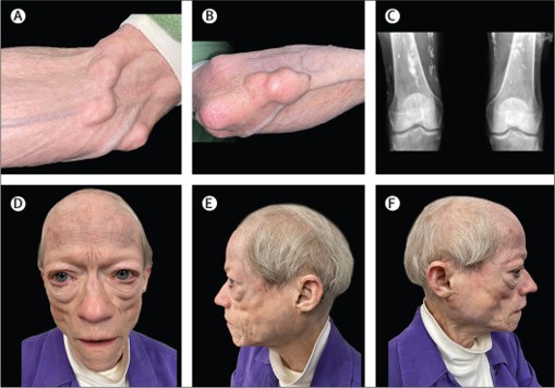
Figure(1): (A and B) Firm subcutaneous nodules (calcinosis cutis) on the arms and prominent superficial veins due to loss of subcutaneous adipose tissue. (C) Radiograph showing bilateral coarse subcutaneous calcifications in the thighs. (D–F) Symmetrical loss of subcutaneous fat (lipodystrophy) from the face (front, left, and right views).
(2). Severe Plasmodium falciparum Malaria
Emilie Karoline Van Pee,Published August 19, 2026,N Engl J Med 2026;395:803,DOI: 10.1056/NEJMicm2603855,VOL. 395 NO. 8
Abstract
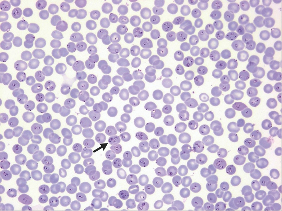
An 82-year-old man was brought to the emergency department after being “found down.” One week earlier, he had returned from a 1-month visit to Chad, his country of birth, and had started having weakness. His body temperature was 37.4°C, and his blood pressure was 88/47 mm Hg. The physical examination was notable for confusion. Laboratory studies showed a hemoglobin level of 12 g per deciliter (reference range, 13.5 to 17), an elevated lactate dehydrogenase level, thrombocytopenia, metabolic acidosis, and acute kidney injury. A peripheral-blood smear showed ring-form trophozoites within erythrocytes and an estimated parasitemia level (expressed as a percentage of erythrocytes) of 50.7% (May–Grünwald Giemsa staining). Some erythrocytes had multiple ring forms (arrow) — a finding highly characteristic of infection with Plasmodium falciparum. Polymerase-chain-reaction testing of the blood was positive for P. falciparum. On the basis of the presence of several criteria — including impaired consciousness, acidosis, renal impairment, and a parasitemia level of greater than 10% — a diagnosis of severe P. falciparum malaria was made. Treatment with intravenous artesunate was initiated and then changed to oral artemisinin-based therapy on hospital day 7, when the parasitemia level was less than 0.5%. On hospital day 8, the hemoglobin level decreased to 5.6 g per deciliter; the decrease was attributed to delayed hemolysis from intravenous artesunate. The patient was discharged home on hospital day 23 in good condition.
(3). Drug-Induced Subacute Cutaneous Lupus Erythematosus
Norbertus A. Ipenburg, Published August 15, 2026,N Engl J Med 2026;395: e10,DOI: 10.1056/NEJMicm2605381,VOL. 395 NO. 8
Abstract
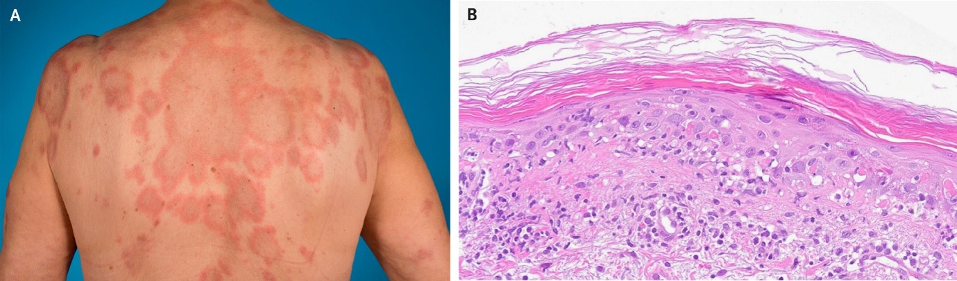
A 73-year-old woman with rheumatoid arthritis presented to the dermatology clinic with burning skin lesions that had emerged on her torso several months earlier. She had taken etanercept for 15 years before the rash started and had been taking pantoprazole for 10 months before the current presentation. Physical examination showed annular polycyclic lesions with indurated, erythematous borders and central clearing on the upper back and arms (Panel A). A quantitative antinuclear antibody test performed with indirect immunofluorescence was positive, and the anti-Ro antibody level was greater than 240 U per milliliter (reference value, <7). Histopathological analysis of a skin-biopsy sample from the patient’s back showed vacuolar interface dermatitis with apoptotic keratinocytes (Panel B, hematoxylin and eosin staining), and Alcian blue staining showed dermal mucin. A diagnosis of drug-induced subacute cutaneous lupus erythematosus was made. Lesions in subacute cutaneous lupus erythematosus have annular and papulosquamous morphologic features and anti-Ro positivity in most cases. A thorough medication history is necessary to differentiate drug-induced from idiopathic subacute cutaneous lupus erythematosus. Culprit medications include thiazides, anti–tumor necrosis factor agents, and proton-pump inhibitors — the last of which was considered to be the trigger in this case. Cessation of the offending drug typically leads to resolution of the rash. Pantoprazole was discontinued, and treatment with hydroxychloroquine and topical clobetasol was started. The rash abated after 4 months and had not recurred 1 year after discontinuation of hydroxychloroquine and topical clobetasol.
(4). Jaccoud’s Arthropathy
Ranjan Gupta, Published August 19, 2026,N Engl J Med 2026;395: e11,DOI: 10.1056/NEJMicm2606133,VOL. 395 NO. 8
Abstract
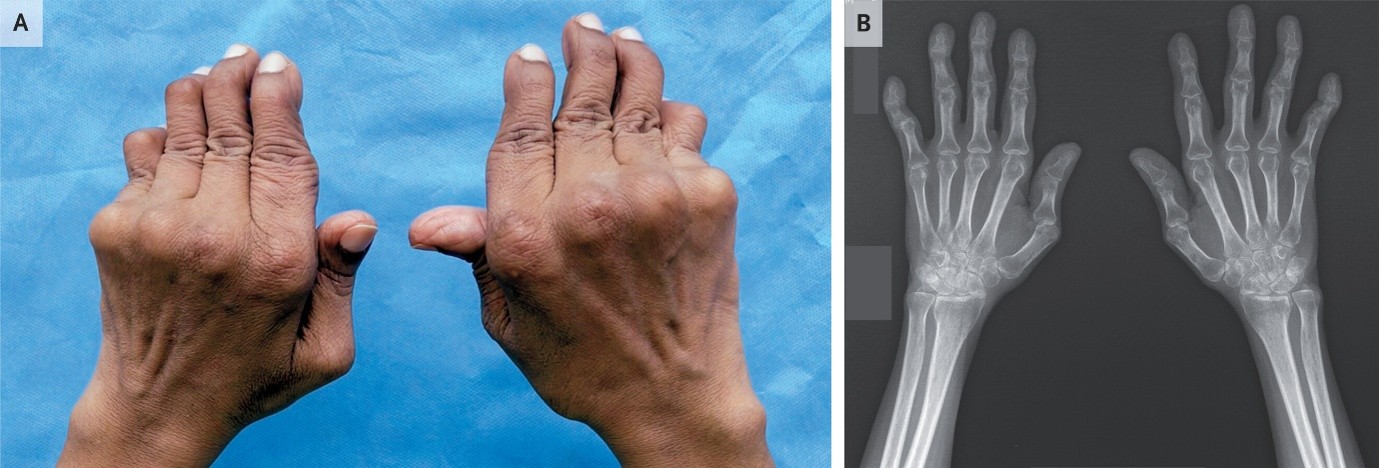
A 30-year-old woman presented to the rheumatology clinic with a 2-year history of pain, swelling, and morning stiffness in the wrists, knuckles, and fingers. During the 6 months before presentation, she had received treatment with methotrexate for possible rheumatoid arthritis, but her symptoms had not abated. Physical examination was notable for swan-neck deformities and slight ulnar deviations of fingers two through five and Z-shaped deformities of the thumbs on both hands when the fingers were extended (Panel A). When the hands were clenched into fists or pressed against the table, the joint deformities largely resolved (Video) — a finding consistent with Jaccoud’s arthropathy. Jaccoud’s arthropathy is a chronic, deforming arthropathy that results from laxity of the ligaments and tendons around joints rather than destruction of bone and cartilage. The deformities are reducible with pressure, and radiography typically does not show bony erosions. Jaccoud’s arthropathy mimics rheumatoid arthritis and is most often associated with systemic lupus erythematosus (SLE). Radiography of the hands showed juxtaarticular osteopenia but no bony erosions (Panel B; labeling has been masked). On the basis of further history taking and serologic testing, a final diagnosis of Jaccoud’s arthopathy with underlying SLE was made. Treatment with hydroxychloroquine, prednisolone, and a higher dose of methotrexate was initiated. At the 4-month follow-up, the patient’s joint pain had abated but the deformities persisted.
(5).Pulmonary Nodules
Matthew E.J. Callister, Published September 2, 2026,N Engl J Med 2026;395:894-905
DOI: 10.1056/NEJMcp2515063,VOL. 395 NO. 9
Summary
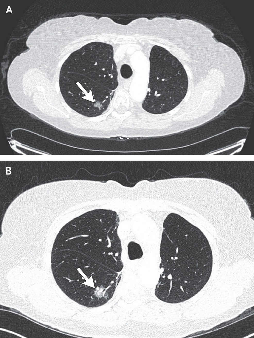
Pulmonary nodules are classified as solid or subsolid, with subsolid nodules further classified as part-solid or pure ground-glass. Management strategies differ according to the type of nodule. A comparison of current and previous imaging studies, when available, is essential to assess the risk of the nodule being malignant. Solid nodules that have been stable for 2 years are considered to be benign, whereas subsolid nodules require a longer period of stability to be considered benign. Prediction models to stratify the risk of the nodule being malignant can be used to guide the management of solid nodules. Computed tomographic (CT) surveillance is indicated for low-risk nodules; positron-emission tomography–CT, biopsy, or both for intermediate-risk nodules; and surgical resection for selected high-risk nodules. Subsolid nodules are often slower growing than solid nodules but are associated with a higher risk of being malignant, especially if a solid component develops or progressively enlarges. Biopsy methods include transthoracic needle biopsy and navigational bronchoscopy. Optimal overall management balances timely diagnosis in persons who have cancer with the avoidance of unnecessary invasive procedures in persons who have benign disease.
(6). Acro-Osteolysis
Nawarat Pengpong, Published September 2, 2026,N Engl J Med 2026;395: e12,DOI: 10.1056/NEJMicm2605116,VOL. 395 NO. 9
Abstract
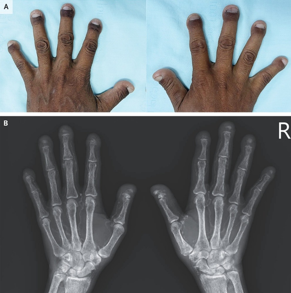
A 46-year-old man with chronic kidney disease presented to the internal medicine clinic with a 2-year history of painless shortening and rounding of the fingertips. Ten years earlier, he had received a diagnosis of chronic kidney disease due to obstructive nephropathy from nephrolithiasis. Since then, he had been intermittently lost to follow-up. On physical examination, all the fingertips were short, broad, and bulbous, with a pseudoclubbing appearance (Panel A). The toes had similar but less severe changes. Radiographs of the hands showed resorption of the terminal tufts of all the distal phalanges (Panel B). The patient was also found to have osteopenia, vascular calcification, chondrocalcinosis of the wrists, and osteolytic lesions in the distal ends of several metacarpals that were consistent with osteitis fibrosa cystica. Bone resorption of the distal phalanges of the hands or feet — known as acro-osteolysis — is associated with several conditions, including systemic sclerosis, Raynaud’s phenomenon, exposure to vinyl chloride, and thermal injury. In this case, renal osteodystrophy and tertiary hyperparathyroidism in the context of end-stage kidney disease were identified as the etiologic factors. Owing to persistent hypercalcemia despite hemodialysis and medical management of hyperparathyroidism, total parathyroidectomy with autotransplantation was performed 3 months after presentation. At a postoperative follow-up visit 6 months after surgery, the patient’s calcium and parathyroid hormone levels had normalized but the fingertips remained unchanged.
(7). Urticarial Vasculitis
Arjun Mahajan.Published September 2, 2026,N Engl J Med 2026;395: e13,DOI: 10.1056/NEJMicm2602968,VOL. 395 NO. 9
Abstract
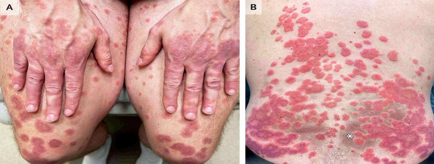
A 33-year-old man with systemic lupus erythematosus (SLE) presented to the dermatology clinic with a 1-week history of pruritic, burning plaques. Two years earlier, he had received a diagnosis of SLE, but treatment had not been started owing to mild symptoms and the absence of end-organ damage. Physical examination was notable for pink, edematous plaques on the hands, elbows, knees, and feet. Laboratory testing showed elevated antinuclear and anti–double-stranded DNA antibody titers and low complement levels. A skin-biopsy sample from the left elbow showed papillary dermal edema, intravascular neutrophils, and C3 deposition in dermal vessel walls — findings consistent with leukocytoclastic vasculitis. Treatment with oral and topical glucocorticoids was started. One week later, the lesions evolved into fixed, urticarial, annular, and targetoid plaques with rims of purpura on the thighs (Panel A), chest, abdomen, and back (Panel B), where hyperpigmentation at the site of healed lesions was also observed (asterisk). On the basis of the presence of urticarial lesions lasting more than 24 hours plus leukocytoclastic vasculitis on histopathological analysis, a diagnosis of urticarial vasculitis was made. Urticarial vasculitis may be associated with SLE (as in this patient), particularly when serum complement levels are low. Treatment with dapsone, colchicine, and hydroxychloroquine was initiated, with eventual replacement of prednisone by mycophenolate mofetil. At a follow-up visit 2.5 years after the start of treatment, the patient’s disease was well controlled.
(8). Severe Plasmodium falciparum Malaria
Emilie Karoline Van Pee, ,Published August 19, 2026,N Engl J Med 2026;395:803,DOI: 10.1056/NEJMicm2603855,VOL. 395 NO. 8.
Abstract
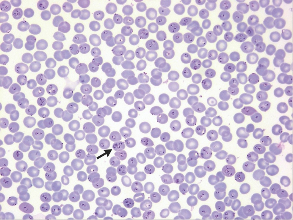
An 82-year-old man was brought to the ED after being “found down.” One week earlier, he had returned from a 1-month visit to Chad. A peripheral-blood smear showed ring-form trophozoites within erythrocytes.
(9). Scrofuloderma
Ana Rita Ambrosio, Published August 29, 2026,DOI: 10.1056/NEJMicm2517515
Abstract
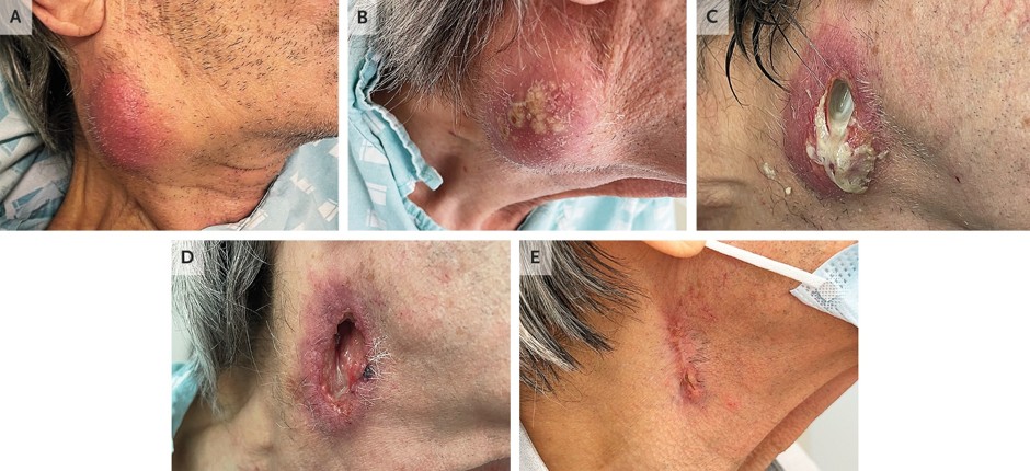
A 72-year-old man presented to the emergency department with a 2-month history of an enlarging, painful lump on his neck. On physical examination, there was a fixed, erythematous, 5-cm mass in the anterior triangle of the neck on the right side (Panel A). Computed tomography (CT) of the neck showed a large necrotic right cervical mass with no identifiable primary tumor. Testing for human immunodeficiency virus was negative, as was an interferon-gamma release assay. The neck mass subsequently enlarged (Panel B, 3 days after presentation). Cytologic analysis of a fine-needle aspiration specimen (obtained 12 days after admission) was consistent with suppurative lymphadenopathy; mycobacterial cultures were negative. The mass subsequently ulcerated and released purulence (Panel C, 17 days after presentation), and a fistulous tract developed (Panel D, 32 days after presentation). A repeat mycobacterial culture of the purulent discharge grew Mycobacterium tuberculosis complex; species-level identification was not performed. A diagnosis of scrofuloderma was made. Scrofuloderma is a form of cutaneous tuberculosis that results from contiguous spread of infection from a deep structure — most commonly a cervical lymph node — into the overlying skin. Antituberculous therapy and wound care were initiated. Evaluation for concurrent pulmonary tuberculosis included chest CT, which showed bilateral pulmonary micronodules that remained stable on follow-up imaging, and bronchoscopy with bronchial sampling. The results of smear microscopy for acid-fast bacilli, nucleic acid amplification testing for M. tuberculosis complex, and mycobacterial cultures (Lowenstein–Jensen and Mycobacterial Growth Indicator Tube) were all negative. Six months after the initiation of treatment, the fistula had healed (Panel E).
(10). Adie Tonic Pupil
Aaron Donnelly, , NEJM, September 5, 2026

Abstract
A 36-year-old man presented to the ophthalmology emergency department with a 1-week history of asymptomatic anisocoria. Physical
examination revealed that the right pupil was dilated (Panel A) and constricted minimally to light and the left pupil constricted briskly to light (Video 1A).
With accommodation, both pupils constricted and the right pupil sustained constriction. Visual acuity, extraocular movements, and deep-tendon reflexes were normal. Administration of dilute pilocarpine (0.125% concentration) to both eyes resulted in more constriction of the right pupil than the left pupil (Panel B), a finding
that indicated denervation hypersensitivity in the right eye. The glycated hemoglobin level was normal. A diagnosis of Adie tonic pupil was made. Adie tonic pupil
is an idiopathic pupillary disorder that is caused by postganglionic parasympathetic denervation of the iris sphincter. It results in light–near dissociation, tonic constriction with accommodation, and cholinergic hypersensitivity in the denervated pupillary muscle. On slit-lamp examination, segmental vermiform movements of the iris due to uneven denervation and partial reinnervation of the iris sphincter muscle were seen (Video 1B, right eye).
Reassurance regarding the benign nature of the diagnosis was provided. At a 3-week follow-up visit, the anisocoria remained
(11). Grey Turner’s Sign
Kan-Ru Li, et al, Published September 9, 2026,N Engl J Med 2026;395:1009,DOI: 10.1056/NEJMicm2605791,VOL. 395 NO. 10
Abstract
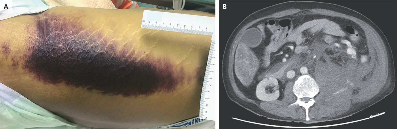
A 46-year-old man with cirrhosis presented to the emergency department with a 1-week history of left flank pain. He reported no recent trauma, medical procedures, or use of anticoagulant or antiplatelet medications. His heart rate was 128 beats per minute, and his blood pressure was 64/37 mm Hg. Physical examination was notable for a large ecchymosis on the left flank — a finding known as Grey Turner’s sign — that was tender to palpation (Panel A; ruler branding has been masked). Grey Turner’s sign may be seen in cases of intraabdominal or retroperitoneal bleeding. Although this finding is classically associated with pancreatitis, it may occur in other conditions, and abdominal imaging is required to identify the underlying cause. Laboratory studies were notable for anemia, severe thrombocytopenia, and metabolic values that yielded a Model for End-Stage Liver Disease score of 26, which indicates advanced liver disease with a substantial risk of death in the short term. Point-of-care ultrasonography of the abdomen showed no intraabdominal free fluid. Computed tomography of the abdomen with administration of contrast material showed active extravasation of the contrast material into the left psoas muscle and retroperitoneal space (Panel B, axial view). A diagnosis of spontaneous retroperitoneal hemorrhage due to coagulopathy of liver disease was made. Transarterial angioembolization of the bleeding vessel was performed, and the bleeding was controlled. After a 3-week hospital stay, the patient was discharged home.–
(12). Late-Onset Pompe Disease
Sofia S. Rodrigues, Published September 16, 2026N Engl J Med 2026;395:1112DOI: 10.1056/NEJMicm2516626VOL. 395 NO. 11A 54
Abstract
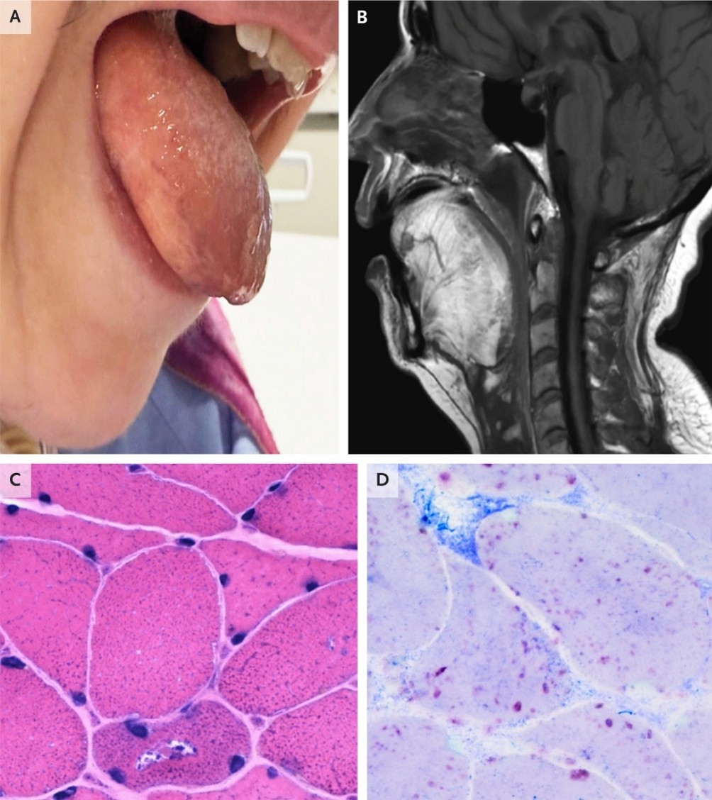
An old woman presented to the emergency department with a 4-month history of progressively worsening asthenia and falls and 5 days of dyspnea. Physical examination was notable for macroglossia (Panel A), head drop, and mild symmetric weakness of the proximal upper and lower limbs. The creatine kinase level was 373 U per liter (reference range, 24 to 173). Magnetic resonance imaging of the head and cervical spine was notable for fatty infiltration of the tongue (Panel B) and paravertebral muscles. Owing to clinical suspicion of an underlying metabolic myopathy, biopsy of the deltoid muscle was performed while results of enzymatic and genetic testing were pending. Hematoxylin and eosin staining of skeletal muscle (Panel C) showed vacuolar myopathy, and acid phosphatase staining (Panel D) showed increased acid phosphatase activity suggestive of lysosomal dysfunction. Enzymatic activity of acid α-glucosidase in leukocytes was low, and genetic testing identified compound heterozygosity for pathogenic variants in GAA. A diagnosis of late-onset Pompe disease, an autosomal recessive lysosomal disorder that manifests as a skeletal myopathy, was made. Treatment with avalglucosidase alfa, an enzyme-replacement therapy, was administered while respiratory failure from ventilatory-muscle weakness and pneumonia was managed. The patient was discharged to an inpatient rehabilitation facility 2 months after presentation.