
Hypertensive intracranial haemorrhage with extensive intraventricular extension in a patient with chronic kidney disease on maintenance haemodialysis: A case report
Jyotiba Bheemappa Hullur1*, Gowdham Pannirselvam2
1Nursing Officer, Kauvery Hospital, Marathahalli, Bengaluru
2Sr. Nurse Educator, Kauvery Hospital, Marathahalli, Bengaluru
*Correspondence
Abstract
Background
Hypertensive intracranial hemorrhage is a life-threatening neurological emergency that can result in rapid neurological deterioration and death. The presence of intraventricular hemorrhage further increases the severity of the condition because it can interfere with cerebrospinal fluid circulation and result in ventricular dilatation and increased intracranial pressure. Patients with advanced chronic kidney disease receiving maintenance hemodialysis represent a high-risk population because of the presence of multiple vascular and systemic comorbidities. This case highlights the clinical progression of severe hypertensive intracranial hemorrhage with extensive intraventricular involvement in a patient with chronic kidney disease on maintenance hemodialysis.
Case Presentation
A 50-year-old female, a known case of chronic kidney disease stage 5D on maintenance hemodialysis, type 2 diabetes mellitus, hypertension and hypothyroidism, presented to the Emergency Department with sudden onset of unresponsiveness. On presentation, her blood pressure was markedly elevated at approximately 220/110 mmHg. Her Glasgow Coma Scale was E1V1M3, and both pupils were approximately 2 mm and non-reactive to light. The patient was intubated and initiated on mechanical ventilation for airway protection. CT brain revealed a left thalamic intracranial hemorrhage measuring approximately 26 × 23 mm with extensive intraventricular extension involving the bilateral lateral, third and fourth ventricles. Ventricular dilatation, effacement of the third ventricle and approximately 4 mm midline shift towards the right were also documented. The patient was managed in the intensive care unit with supportive and neuroprotective measures, anti-epileptic therapy and multidisciplinary management. An external ventricular drain was inserted because of the intraventricular hemorrhage and ventricular changes. Despite continued intensive and neurosurgical management, the patient’s neurological condition progressively deteriorated. Absence of brainstem reflexes was subsequently documented. The family was counselled regarding the patient’s poor prognosis and opted for treatment in accordance with comfort measures. On 11 July 2026, the patient developed cardiac arrest. Resuscitative measures were initiated according to the ACLS protocol; however, despite continued efforts, the patient could not be revived and was declared deceased at 10:45 PM. The documented causes of death were hypertensive intracranial hemorrhage, neurogenic shock and brainstem death.
Conclusion
This case demonstrates the severe clinical course of hypertensive intracranial hemorrhage with extensive intraventricular extension in a patient with advanced chronic kidney disease on maintenance hemodialysis. Early recognition of neurological deterioration, prompt neuroimaging, appropriate airway management, continuous neurological monitoring and timely neurosurgical intervention are essential in such critically ill patients. The case also highlights the importance of multidisciplinary management, meticulous intensive care nursing and clear communication with the family when the neurological prognosis becomes poor.
Keywords: Hypertensive intracranial hemorrhage; Thalamic hemorrhage; Intraventricular hemorrhage; chronic kidney disease; Hemodialysis; External ventricular drain; Brainstem death; Intensive care nursing.
Introduction
Intracerebral hemorrhage is a neurological emergency caused by rupture of a blood vessel within the brain parenchyma. It can produce direct tissue injury, mass effect, increased intracranial pressure and secondary neurological damage. Among the different causes of spontaneous intracerebral hemorrhage, chronic hypertension is one of the most important risk factors, particularly for hemorrhages occurring in deep structures such as the basal ganglia, thalamus, brainstem and cerebellum. The outcome depends on several factors, including the location and size of the hematoma, level of consciousness at presentation, presence of intraventricular hemorrhage, hydrocephalus, age and associated medical conditions.
Intraventricular hemorrhage refers to the extension of blood into the ventricular system. It can occur as a consequence of an intracerebral hemorrhage and is clinically important because blood within the ventricles can interfere with normal cerebrospinal fluid circulation. This can lead to acute hydrocephalus and increased intracranial pressure. Hydrocephalus following intracerebral hemorrhage is associated with poor outcomes, and ventricular drainage may be required in selected patients. Current guidelines recommend ventricular drainage in patients with intracerebral hemorrhage or intraventricular hemorrhage when hydrocephalus is contributing to a reduced level of consciousness.
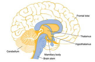
Image Source: Pubmed Central; The Role of Thiamine Deficiency in Alcoholic Brain Disease; Peter R Martin et al, 2003;27(2)
Chronic kidney disease is also strongly associated with cerebrovascular disease. Patients with advanced chronic kidney disease and end-stage kidney disease have increased risks of both ischemic and haemorrhagic stroke. Multiple factors may contribute, including hypertension, diabetes, vascular calcification, endothelial dysfunction, inflammation, altered platelet function and abnormalities in coagulation. Patients undergoing hemodialysis represent a particularly vulnerable population.
Anatomy and Physiology
Brain
The brain is the principal organ of the central nervous system and is responsible for controlling and coordinating a wide range of functions, including consciousness, movement, sensation, cognition, speech, memory, autonomic regulation and several endocrine functions. The cerebral blood supply is primarily derived from the internal carotid and vertebrobasilar arterial systems. These vessels form an interconnected arterial network and give rise to branches that supply both superficial and deep regions of the brain. Small penetrating arteries supply structures located deep within the cerebral hemispheres. These small vessels are particularly susceptible to chronic hypertensive vascular changes. When a cerebral blood vessel ruptures, blood accumulates within the surrounding brain tissue. The resulting hematoma produces direct mechanical injury to neurons and surrounding structures. Because the skull is a fixed compartment, accumulation of blood also occupies intracranial space and may increase intracranial pressure. If the hemorrhage is large or associated with ventricular obstruction, cerebral perfusion can become compromised, resulting in secondary brain injury.
Thalamus
The thalamus is a paired deep gray matter structure located within the diencephalon. It lies on either side of the third ventricle and has close anatomical relationships with several important structures of the brain. The thalamus functions mainly as a relay and integration centre for sensory and motor information. It also has an important role in maintaining consciousness, alertness, sleep and communication between different regions of the brain. Because of its deep location and vascular supply from small penetrating arteries, the thalamus can be involved in hypertensive intracerebral hemorrhage. A hemorrhage involving the thalamus can produce a significant neurological deficit depending on the size and direction of hematoma extension. Involvement of nearby ventricular structures is particularly important because the thalamus is closely related to the third ventricle. In the present case, CT brain demonstrated a left thalamic hematoma measuring approximately 26 × 23 mm. The hemorrhage extended into the ventricular system, making the clinical condition more severe.
Ventricular System
The ventricular system consists of the paired lateral ventricles, third ventricle, cerebral aqueduct and fourth ventricle. These interconnected cavities contain cerebrospinal fluid. Cerebrospinal fluid provides mechanical protection to the brain and spinal cord, contributes to maintenance of the intracranial environment and participates in the removal of metabolic waste. CSF is produced mainly by the choroid plexus and circulates through the ventricular system before reaching the subarachnoid space and eventually being reabsorbed into the venous circulation. Under normal conditions, CSF production and absorption remain balanced. When blood enters the ventricular system, this normal circulation can become disturbed. Blood products may obstruct CSF pathways and interfere with CSF absorption. The resulting accumulation of CSF can produce ventricular enlargement and hydrocephalus. In this patient, blood was reported within the bilateral lateral ventricles, third ventricle and fourth ventricle. The imaging also demonstrated ventricular dilatation and periventricular CSF leakage, indicating significant disturbance of normal CSF dynamics.
Pathophysiology
The pathophysiology of hypertensive intracerebral hemorrhage begins with long-term structural damage to small cerebral vessels. Persistent hypertension produces changes in the vessel walls and makes them vulnerable to rupture. When the pressure within the vessel exceeds the strength of the damaged vascular wall, the vessel may rupture and blood enters the brain parenchyma. In the present case, severe hypertension was documented at presentation. The initial systolic blood pressure was approximately 200 mmHg, with subsequent readings reaching 220/110 mmHg. The hemorrhage occurred in the left thalamic region, which is a recognized location for deep hypertensive hemorrhage. The initial hematoma produces direct injury to the surrounding neuronal tissue. As the hematoma increases or extends, it can compress adjacent structures and increase intracranial pressure. In this patient, the hemorrhage extended from the thalamic region into the ventricular system. Once blood entered the ventricles, the normal circulation of CSF was affected. The presence of blood within the bilateral lateral, third and fourth ventricles contributed to ventricular dilatation. The CT findings also described effacement of the third ventricle and a mild midline shift of approximately 4 mm towards the right.
The development of ventricular dilatation can further increase intracranial pressure. Increased intracranial pressure can reduce cerebral perfusion and cause secondary brain injury. If the process continues, progressive dysfunction of deeper neurological structures and eventually brainstem dysfunction may occur. Intraventricular hemorrhage is recognized as an important adverse prognostic factor in intracerebral hemorrhage. Earlier literature and contemporary reviews have consistently associated IVH with increased risk of hydrocephalus and poor neurological outcomes.
Chronic Kidney Disease and Cerebrovascular Risk
The patient’s underlying chronic kidney disease stage 5D and dependence on maintenance hemodialysis were important clinical considerations. Chronic kidney disease is not only a renal disorder but is associated with systemic vascular and hematological changes. Patients with advanced CKD have increased risk of cerebrovascular disease due to the combination of traditional cardiovascular risk factors and mechanisms related specifically to renal dysfunction.
Hypertension and diabetes are common in patients with advanced kidney disease and are themselves major vascular risk factors. In addition, CKD can be associated with endothelial dysfunction, chronic inflammation, vascular calcification and abnormalities in platelet and coagulation function. Uremic toxins and changes in cerebral vascular regulation may also contribute to cerebrovascular vulnerability.
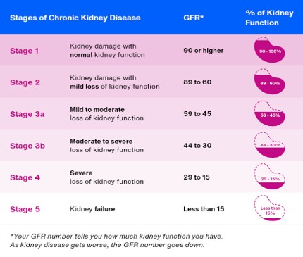
Image Source: kidney.org; Kidney Failure Risk Factor: Estimated Glomerular Filtration Rate (eGFR)
Patients receiving hemodialysis represent a particularly high-risk group. Previous studies have reported increased incidence of stroke among dialysis patients, with hemorrhagic stroke accounting for a substantial proportion of events. In one clinical series of patients with intracerebral hemorrhage, patients receiving hemodialysis had higher rates of intraventricular hemorrhage and higher mortality than patients not receiving hemodialysis. More recent evidence also supports the association between CKD and poor outcomes following intracerebral hemorrhage. A 2026 meta-analysis involving approximately 5,000 patients found that CKD was associated with higher short-term and one-year mortality, greater functional dependence and increased risk of hematoma expansion, with the greatest risks among patients with severe CKD or dialysis dependence.
In the present patient, the coexistence of CKD stage 5D, maintenance hemodialysis, hypertension and diabetes represented a significant background of vascular risk. However, because the available death summary does not provide detailed laboratory findings, dialysis timing or anticoagulation details, a direct causal relationship between any specific dialysis-related factor and the hemorrhage cannot be established from this case.
Case Presentation
A 50-year-old female presented to the Emergency Department, with sudden onset of unresponsiveness. She was a known case of type 2 diabetes mellitus, hypertension, hypothyroidism and chronic kidney disease stage 5D on maintenance hemodialysis. According to the clinical documentation, the patient had a history of hypertension and was using medication on an SOS basis when her systolic blood pressure was elevated. A blood pressure measurement approximately one week before the present event had been documented, and a blood pressure of 140/80 mmHg was recorded on 05/07/2026. However, at the time of the acute presentation, the patient’s systolic blood pressure was markedly elevated.
On arrival at the Emergency Department, her blood pressure was documented as approximately 220/110 mmHg, with subsequent readings ranging from 171/96 mmHg onwards. The patient was unresponsive and had a very low GCS score of E1V1M3. Both pupils were approximately 2 mm and non-reactive to light. In view of the low level of consciousness and inability to adequately protect the airway, the patient was intubated. Mechanical ventilation was initiated for airway protection and respiratory support. The patient was subsequently shifted to the Medical Intensive Care Unit for further evaluation and management.
Radiological Findings
An emergency CT brain was performed following the patient’s presentation. The scan demonstrated an intracranial hemorrhage involving the left thalamus, measuring approximately 26 × 23 mm. The hemorrhage extended into the ventricular system, involving the bilateral lateral ventricles, third ventricle and fourth ventricle. The reported imaging findings also included effacement of the third ventricle and approximately 4 mm of midline shift towards the right. Ventricular dilatation with periventricular CSF seepage was also described. The combination of a deep thalamic hematoma, extensive intraventricular extension and ventricular dilatation indicated a severe intracranial event with a high risk of secondary neurological deterioration.
Initial Management
Following stabilization in the Emergency Department, the patient was transferred to the Medical Intensive Care Unit for further management. Mechanical ventilation was continued because of the patient’s poor level of consciousness. Neurological status was monitored closely, along with other supportive intensive care parameters. Anti-epileptic therapy and other supportive and neuroprotective measures were provided during the hospital and ICU stay. The patient was evaluated by the relevant multidisciplinary teams, including neurology and neurosurgery, because of the extensive intracranial and intraventricular involvement. Because the CT findings demonstrated intraventricular hemorrhage with ventricular dilatation, neurosurgical intervention was considered necessary.
External Ventricular Drainage
An external ventricular drain was inserted on 06/07/2026 under general anesthesia. An EVD provides temporary drainage of cerebrospinal fluid from the ventricular system and can be used to manage acute hydrocephalus and elevated intracranial pressure associated with intraventricular hemorrhage. The American Heart Association/American Stroke Association guideline recommends ventricular drainage in patients with intracerebral or intraventricular hemorrhage when hydrocephalus is contributing to reduced consciousness.
In this patient, the EVD was inserted as part of the management of the ventricular involvement. Despite this intervention and continued intensive care, the patient’s neurological condition did not improve.
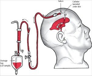
Image Source: Neupsy Key, External Ventricular Drain
Further Clinical Course
During the subsequent ICU stay, the patient continued to receive supportive treatment and neurological monitoring. A repeat CT brain was performed to assess the progression of the intracranial hemorrhage. The repeat scan showed an interval increase in the size of the left thalamic hematoma with increased intraventricular extension and worsening ventricular changes. The imaging also demonstrated increased ventricular dilatation and periventricular CSF seepage. The patient continued to have poor neurological status despite aggressive medical and neurosurgical management. Anti-epileptic therapy and other supportive measures were continued. The clinical condition was reviewed by the neurology and neurosurgery teams during the ICU stay. A CSF analysis was also performed during the hospital course. A further CT brain was performed on 07/07/2026 following placement of the external ventricular drain. The imaging continued to demonstrate significant intraventricular involvement and ventricular changes. The patient remained critically ill despite the interventions. Her neurological status continued to deteriorate over the following days.
Neurological Deterioration
On subsequent assessment, the patient’s neurological condition remained extremely poor. On 08/07/2026, the patient clinical condition indicated further worsening of the neurological status. By 09/07/2026, her condition had deteriorated further with absence of brainstem reflexes.
Final Event and Outcome
On 11/07/2026 at approximately 10:15 PM, the patient developed an episode of cardiac arrest. Resuscitative measures were initiated according to the Advanced Cardiovascular Life Support protocol. Despite the best efforts of the treating team, return of spontaneous circulation could not be achieved. The patient was declared deceased on 11/07/2026 at 10:45 PM.
The documented cause of death was hypertensive intracranial hemorrhage, neurogenic shock and brainstem death. The secondary diagnoses were chronic kidney disease stage 5D on maintenance hemodialysis, type 2 diabetes mellitus, hypertension and hypothyroidism.
Nursing Management and Clinical Significance
The nursing role in this case was particularly important because the patient required continuous intensive care and neurological monitoring. In patients with severe intracranial hemorrhage, frequent assessment of the level of consciousness, Glasgow Coma Scale, pupillary size and reaction, motor response and other neurological parameters is necessary to identify changes in neurological status. Any sudden deterioration should be immediately communicated to the treating team. Airway management was another major component of care. Since the patient presented with severely depressed consciousness and was unable to protect her airway, endotracheal intubation and mechanical ventilation were required. Nursing care included monitoring of the endotracheal tube, ventilator parameters, oxygenation, secretion management and prevention of complications associated with prolonged mechanical ventilation. Management of the external ventricular drain required strict adherence to the prescribed level and drainage system. The nursing team must ensure that the drainage system remains secure and appropriately positioned and should observe the amount and characteristics of CSF drainage. Sudden changes in drainage, obstruction, accidental disconnection, leakage or changes in neurological status require immediate attention. Strict aseptic technique is essential because EVD-associated infection can result in serious complications. Fluid balance was also important because the patient had chronic kidney disease stage 5D and was receiving maintenance hemodialysis. Accurate intake and output monitoring, assessment of fluid status and coordination with the nephrology team are important in such patients. However, specific dialysis parameters and laboratory results were not available in the death summary and therefore have not been included in this report. The nursing team also had an important role in preventing secondary complications associated with prolonged ICU stay. Regular repositioning, pressure injury prevention, infection prevention, oral care, eye care, nutritional support as prescribed and maintenance of skin integrity are important aspects of comprehensive critical care. As the patient’s neurological condition deteriorated, communication and psychosocial support became increasingly important. The nursing team participated in maintaining patient dignity, facilitating communication between the family and healthcare team and supporting the family during the transition towards comfort-focused care.
Discussion
The present case represents a severe hypertensive intracerebral hemorrhage in a patient with multiple comorbidities, presenting with sudden unresponsiveness, severe hypertension and a very low GCS of E1V1M3. CT brain revealed a left thalamic hemorrhage with extensive intraventricular extension. Thalamic hemorrhage is a recognized form of deep hypertensive intracerebral hemorrhage because the thalamus is supplied by small penetrating cerebral vessels. The extensive intraventricular extension in this patient involved the bilateral lateral, third and fourth ventricles and was associated with ventricular dilatation. Blood within the ventricular system can interfere with normal CSF circulation and contribute to hydrocephalus and increased intracranial pressure. Due to the ventricular involvement, an external ventricular drain was inserted to facilitate CSF drainage and manage the ventricular changes. Despite EVD placement and continued intensive care, the patient’s neurological condition progressively deteriorated. The patient’s chronic kidney disease stage 5D, maintenance hemodialysis, hypertension and diabetes mellitus added significant complexity to her clinical condition and represented important vascular risk factors. However, based on the available documentation, the hemorrhage cannot be attributed to any single factor. The subsequent absence of brainstem reflexes indicated severe neurological injury. The death summary documents multidisciplinary counselling regarding the poor prognosis and futility of continuing aggressive treatment, following which the family opted for comfort-focused care. The patient later developed cardiac arrest and could not be revived despite resuscitative efforts. This case highlights the importance of early recognition of intracerebral hemorrhage, prompt neuroimaging, continuous neurological monitoring, timely neurosurgical intervention and multidisciplinary management, particularly in patients with advanced CKD and multiple comorbidities.
Normal Versus Abnormal Process in This Case
Under normal conditions, cerebral blood vessels maintain a regulated blood supply to the brain, while the ventricular system continuously produces and circulates CSF. Cerebral perfusion and intracranial pressure remain within a physiological range that supports normal neuronal function. In this patient, severe hypertension was associated with intracranial vessel rupture and formation of a left thalamic hematoma. The blood subsequently extended into the ventricular system, disturbing normal CSF circulation. Ventricular enlargement developed and the third ventricle was described as effaced. The resulting intracranial changes were associated with severe impairment of consciousness and progressive neurological deterioration. The normal process can therefore be represented as stable cerebral circulation → intact cerebral vessels → normal CSF production and circulation → stable intracranial pressure → preserved neurological function.
The abnormal process in this case progressed as severe hypertension → vascular injury → vessel rupture → thalamic hemorrhage → intraventricular extension → disturbed CSF circulation → ventricular dilatation → increased intracranial pressure and secondary brain injury → neurological deterioration → brainstem dysfunction.
Conclusion
This case describes a severe hypertensive intracranial hemorrhage involving the left thalamus with extensive intraventricular extension in a 50-year-old female with chronic kidney disease stage 5D on maintenance hemodialysis, type 2 diabetes mellitus, hypertension and hypothyroidism. The patient presented with sudden unresponsiveness, severe hypertension and markedly depressed consciousness. Neuroimaging demonstrated a left thalamic hematoma with extension into the bilateral lateral, third and fourth ventricles, associated with ventricular dilatation and midline shift. Despite early airway protection, mechanical ventilation, intensive care management, anti-epileptic therapy, supportive and neuroprotective measures and external ventricular drainage, the patient’s neurological status progressively deteriorated. The subsequent development of absent brainstem reflexes reflected the severity of the neurological injury. Following multidisciplinary counselling, the family opted for comfort-focused treatment. The patient later developed cardiac arrest and was declared deceased despite resuscitative efforts. The case emphasizes the complex interaction between severe hypertension, advanced chronic kidney disease, hemodialysis and intracerebral hemorrhage. It also demonstrates the importance of early recognition, prompt neuroimaging, neurological monitoring, appropriate neurosurgical intervention, meticulous ICU nursing care and effective communication with the family in patients with catastrophic intracranial hemorrhage.
References
- Greenberg SM, Ziai WC, Cordonnier C, Dowlatshahi D, Francis B, Goldstein JN, et al. 2022 Guideline for the Management of Patients With Spontaneous Intracerebral Hemorrhage: A Guideline From the American Heart Association/American Stroke Association. Stroke. 2022;53(7):e282-e361. doi:10.1161/STR.0000000000000407.
- Dey M, Jaffe J, Stadnik A, Awad IA. External ventricular drainage for intraventricular hemorrhage. Curr Neurol Neurosci Rep. 2012;12(1):24-33. doi:10.1007/s11910-011-0231-x.
- Kirmani AR, Sarmast AH, Bhat AR. Role of external ventricular drainage in the management of intraventricular hemorrhage; its complications and management. Surg Neurol Int. 2015;6:188. doi:10.4103/2152-7806.172533.
- Haldrup M, Miscov R, Mohamad N, Rasmussen M, Dyrskog S, Simonsen CZ, et al. Treatment of intraventricular hemorrhage with external ventricular drainage and fibrinolysis: a comprehensive systematic review and meta-analysis of complications and outcome. World Neurosurg. 2023;174:183-196.e6. doi:10.1016/j.wneu.2023.01.021.
- Power A. Stroke in dialysis and chronic kidney disease. Blood Purif. 2013;36(3-4):179-183. doi:10.1159/000356086.
- Iseki K, Fukiyama K. Evidence for high risk of cerebral hemorrhage in chronic dialysis patients. Kidney Int. 1993;44(5):1086-1090. doi:10.1038/ki.1993.352.
- Nayak-Rao S, Shenoy MP. Stroke in patients with chronic kidney disease: How do we approach and manage it? Indian J Nephrol. 2017;27(3):167-171. doi:10.4103/0971-4065.202405.
- El Husseini N, Kaskar O, Goldstein LB. Chronic kidney disease and stroke. Adv Chronic Kidney Dis. 2014;21(6):500-508. doi:10.1053/j.ackd.2014.09.001.
- Power A, Chan K, Singh SK, Taube D, Duncan N. Appraising stroke risk in chronic kidney disease. J Am Soc Nephrol. 2012;23(11):1816-1822.
- Greer DM, Kirschen MP, Lewis A, Gronseth GS, Rae-Grant A, Ashwal S, et al. Pediatric and Adult Brain Death/Death by Neurologic Criteria Consensus Guideline. Neurology. 2023;101(24):1112-1132. doi:10.1212/WNL.0000000000207740.
- Lewis A, Kirschen MP, Greer D. The 2023 AAN/AAP/CNS/SCCM Pediatric and Adult Brain Death/Death by Neurologic Criteria Consensus Practice Guideline: A Comparison With the 2010 and 2011 Guidelines. Neurol Clin Pract. 2023;13(6):e200189. doi:10.1212/CPJ.0000000000200189.
- Wu W, Li Q. Mechanisms of hydrocephalus after intraventricular haemorrhage: a review. Childs Nerv Syst. 2025;41(1):49. doi:10.1007/s00381-024-06711-2.